DISRUPT CAD Pooled OCT Subgroup Analysis
Explore the OCT sub-analysis of individual patient data pooled from four coronary studies to see Shockwave Intravascular Lithotripsy (IVL) outcomes, fracture analysis and stent expansion predictors across various calcium arcs and nodule presence.
DISRUPT CAD Pooled OCT Analysis Conclusions
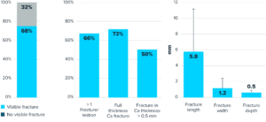
- The present individual patient data pooled analysis of four studies (N=262) represents the largest evaluation of Shockwave IVL by OCT
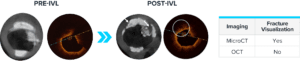
- OCT demonstrated extensive calcium fracture after Shockwave IVL treatment with excellent stent expansion of severely calcified lesions
- Visible calcium fracture and calcium characteristics were not predictors of stent expansion following treatment with Shockwave IVL
Moderated Poster, TCT 120: OCT Characterization of Shockwave IVL for Treatment of Calcified Coronary Lesions: Patient-level Pooled Analysis of the DISRUPT CAD OCT Sub-studies, Ziad A. Ali, TCT 2021.
| DISRUPT CAD I | DISRUPT CAD II | DISRUPT CAD III | DISRUPT CAD IV | DISRUPT CAD Pooled | |
| Enrollment | Dec 2015 – Sep 2016 | May 2018 – Mar 2019 | Jan 2019 – Mar 2020 | Nov 2019 – Apr 2020 | Dec 2015 – Apr 2020 |
| Study Design | Prospective, multi-arm, single-arm | ||||
| ITT (N) | 601 | 1203 | 3844 | 645 | 6286 |
| OCT Analysis* | 282 | 57 | 106* | 71* | 262 |
| OCT Core Laboratory | Cardiovascular Research Foundation, New York, NY | ||||
| Target Lesions | Severely calcified**, de novo coronary artery lesions | ||||
*Patient enrollment in OCT sub-studies was open to all sites participating in the DISRUPT CAD studies that routinely perform OCT imaging.
** Includes patients from DISRUPT CAD III and IV.
1: Brinton et al. Circulation 2019;139:834-836
2: Ali et al. J Am Coll Cardiol Img 2017;10:897-906
3: Ali et al. Circ Cardiovasc Interv 2019;12:e008434
4: Hill et al. J Am Coll Cardiol 2020;76:2635-46
5: Saito et al. Circ J 2021;85(6):826-33
6:Kereiakes et al., J Am Coll Cardiol Intv 2021;14:1337-48
Accordion Section
More DISRUPT CAD Studies
-
Single-arm, pre-market study demonstrating the safety and performance of Shockwave IVL in heavily calcified coronary lesions.Coronary IVL
-
European post-market study confirming DISRUPT CAD I results and showing strong safety and procedural success with Shockwave coronary IVL.Coronary IVL
-
Largest and most rigorous Shockwave study showcasing safety, effectiveness and ease of use of Shockwave coronary IVL.Coronary IVL