Join Drs. Lindsey Cilia and Allen Jeremias as they crack into Shockwave C2 Aero’s aero-volutionary deliverability and crossability during an on-site interview at CRT 2026. Watch as they discuss the benefits of this new device compared to prior generations of Coronary IVL, dive into the ways it’s impacted their calcium algorithms, and walk through a calcified tortuous RCA live case performed by Dr Jeremias and his team.

How Has Shockwave Coronary IVL Changed Your Practice? (0:10 -1:15)

Drs. Cilia and Jeremias talk about how safe, effective Coronary IVL has significantly changed the way physicians tackle complex PCI — helping them improve their outcomes especially in complex, tortuous lesions.

Tell Us About Your Experience with Shockwave C2 Aero (1:15-2:15)

Drs. Cilia and Jeremias discuss Shockwave C2 Aero’s device’s improved deliverability, including how it requires less reliance on adjunctive tools compared to Shockwave C2+.

“A lot of times it’s my first go-to device for calcium modification even if the lesion is super tortuous and the imaging catheter won’t cross.”

—Dr. Lindsey Cilia

“It’s an amazing improvement from the first generation — it’s like going from iPhone 1 to iPhone 15.”

— Dr. Allen Jeremias

How Has Shockwave C2 Aero Impacted Your Treatment Algorithm? (2:20-3:15)

From use as an upfront calcium modification tool to helping enable a radial approach in more complex patients, Drs Cilia and Jeremias cover all the advancements Shockwave C2 Aero has brought to their practices.

“My go-to is really to take this device up front as opposed to some of the more high-risk devices.”

—Dr. Lindsey Cilia

“Many times it avoids using atherectomy … and it shortens the procedure and just simplifies it.”

— Dr. Allen Jeremias

Case Review: PCI of Calcified Tortuous RCA (3:15-end)

Dr. Jeremias walks Dr. Cilia through a recent Shockwave C2 Aero case involving a 74-year-old male patient with multiple comorbidities and a heavy coronary calcium burden involving three different morphologies.

Watch to hear their entire conversation about this complex case, including the importance of imaging, reducing patient time on the table, and how Shockwave C2 Aero’s deliverability, crossability and re-wrapability performed in a real-world scenario.

Explore Shockwave C2 Aero


Dr. Jeremias and Dr. Cilia are paid consultants of Shockwave Medical.

Shockwave IVL: In the U.S.: Rx only. Prior to use, please reference the Important Safety Information for more information on indications, contraindications, warnings, precautions and adverse events.
At SIR 2026, Dr. Narayanan Thulasidasan, Consultant Interventional Radiologist at Guy’s and St Thomas’ NHS Foundation Trust in London, UK, shares four challenging distal tibial and below-the-ankle (BTA) revascularization cases. These case reviews highlight treatment strategies for heavily calcified disease, procedural decision-making, and the use of Shockwave Javelin Peripheral Intravascular Lithotripsy (IVL) to facilitate successful limb salvage interventions.

Case 1: Distal Tibial & Below-the-Ankle Revascularization Using Shockwave Javelin Peripheral IVL

Dr. Narayanan Thulasidasan presents the treatment of a complex below-the-ankle (BTA) chronic limb-threatening ischemia (CLTI) case in an 86-year-old patient with toe necrosis. Faced with heavily calcified distal pedal disease and limited treatment options, the team used Shockwave Javelin Peripheral IVL to facilitate lesion crossing and vessel preparation, followed by IVL and angioplasty. The case highlights decision-making in challenging distal tibial and pedal interventions and demonstrates successful restoration of blood flow to support wound healing.

Case 2: Complex Pedal Arch Revascularization in a High-Risk CLTI Patient

In this video, Dr. Thulasidasan discusses a challenging below-the-ankle intervention in a patient with diabetes, end-stage renal disease, and tissue loss in a remaining limb. With severe calcification limiting device delivery, Shockwave Javelin Peripheral IVL was used to facilitate crossing and treatment of the dorsalis pedis, pedal arch, and posterior tibial artery. The case highlights procedural strategies for navigating complex pedal anatomy, optimizing lesion preparation, and restoring inline flow to support wound healing and limb preservation.

Case 3: Treating Severe Below-the-Ankle Calcification with Shockwave Javelin Peripheral IVL

Dr. Thulasidasan presents a case involving an 86-year-old patient with a non-healing hallux ulcer caused by a nail bed injury. Faced with extensive calcification and difficult catheter delivery below the ankle, the team used Shockwave Javelin Peripheral IVL to modify calcium in the posterior tibial artery and plantar circulation after conventional crossing techniques proved insufficient. The case highlights procedural decision-making, lesion preparation strategies, and the role of Shockwave Javelin Peripheral IVL in facilitating treatment of heavily calcified distal tibial and pedal vessels to improve perfusion and support wound healing.

Case 4: Urgent Limb Salvage Following Partial Foot Amputation

Dr. Thulasidasan presents a complex Rutherford 5 chronic limb-threatening ischemia (CLTI) case in a patient who required emergency partial foot amputation due to infection and sepsis. With severe below-the-ankle calcification limiting conventional treatment options, Shockwave Javelin Peripheral IVL was used to modify calcium in the posterior tibial and plantar arteries, enabling successful balloon delivery and revascularization. The case highlights the role of Shockwave Javelin Peripheral IVL in overcoming heavily calcified distal disease, restoring blood flow after urgent surgical intervention, and supporting ongoing wound healing.


Dr. Thulasidasan is a paid consultant of Shockwave Medical.

Shockwave IVL: In the U.S.: Rx only. Prior to use, please reference the Important Safety Information for more information on indications, contraindications, warnings, precautions and adverse events.
In this case, Dr. Jeff Tyler, MD (Orange County Heart Institute, Director of Cardiogenic Shock at Providence St. Joseph Hospital), takes on a severely stenosed lesion that initially resisted intravascular ultrasound (IVUS) crossing. Shockwave C2 Aero demonstrates its enhanced deliverability and crossability, successfully navigating the lesion where traditional imaging tools could not.

By overcoming these procedural barriers, Shockwave C2 Aero helps simplify an otherwise extremely difficult intervention and enables treatment in a case that would typically present significant limitations.

Watch the case review to learn more.


Dr. Tyler is a paid consultant of Shockwave Medical.

Shockwave IVL: In the U.S.: Rx only. Prior to use, please reference the Important Safety Information for more information on indications, contraindications, warnings, precautions and adverse events.
Curious how Shockwave C2 Aero performs in tough, calcified lesions? In this case review, Dr. Yousif Ahmad and Dr. Brett Wanamaker take you step‑by‑step through an initial Rotashock procedure, demonstrating how Shockwave C2 Aero’s enhanced deliverability and crossability help safely increase vessel compliance before stenting.

Patient Overview: Complex Calcified Left Main Bifurcation

An 85-year-old female patient presented with worsening angina and dyspnea. Angiography showed severely calcified distal left main bifurcation disease, impacting both the left anterior descending artery (LAD) and diagonal branches.

Deploying Shockwave C2 Aero Post-Atherectomy to Increase Compliance Before Stenting

Dr. Ahmad used atherectomy in the LAD and circumflex, resulting in a slow flow complication in the LAD that was resolved with bailout stenting. He followed with a 4.0 mm Shockwave C2 Aero catheter to achieve complete calcium modification. Dr. Ahmad first delivered 60 pulses in the left circumflex artery (LCX), then advanced the catheter easily into the proximal LAD lesion to deliver the remaining 60 pulses. Finally, he placed a stent in the left main bifurcation.

Post-Case Reflection: Next-Gen Capabilities Could Enable Shockwave IVL-First Strategy

Dr. Ahmad and Dr. Wanamaker discuss how Shockwave C2 Aero’s upgrades — increased deliverability, crossability and re-wrap ability — in conjunction with Shockwave’s proven safety and efficacy have the potential to shift workflows to a Shockwave IVL-first approach.


Watch the full case review to learn more about Shockwave C2 Aero’s capabilities in complex coronary interventions.


Dr. Ahmad and Dr. Wanamaker are paid consultants of Shockwave Medical.

Shockwave IVL: In the U.S.: Rx only. Prior to use, please reference the Important Safety Information for more information on indications, contraindications, warnings, precautions and adverse events.
Dr. Brett Wanamaker, MD, FACC, FSCAI, Associate Professor of Internal Medicine at the University of Michigan, presents a Shockwave C2 Aero case demonstrating improved deliverability and crossability in complex calcified coronary lesions.

Treating Complex Calcified LAD and RCA CTO Lesions

This case highlights treatment of a severely calcified left anterior descending artery (LAD) and a right coronary artery (RCA) chronic total occlusion (CTO). The Shockwave C2 Aero successfully crossed a tortuous proximal LAD and the RCA CTO with minimal guidewire or catheter support, demonstrating strong lesion crossing capability in challenging anatomy.

Overcoming Failed Stent Delivery

Although noncompliant balloons were able to cross both lesions, initial stent delivery attempts were unsuccessful—highlighting the limitations of conventional approaches in heavily calcified lesions.

Enabling Stent Delivery with Calcium Modification

Following calcium modification with Shockwave C2 Aero, stent delivery was achieved. Final imaging confirmed strong stent expansion with a high minimal stent area (MSA), supporting a favorable procedural outcome and positive patient prognosis.


Watch the full case review to see how Shockwave C2 Aero improves crossability, enables stent delivery, and optimizes outcomes in complex calcified coronary lesions.


Dr. Wanamaker is a paid consultant of Shockwave Medical.

Shockwave IVL: In the U.S.: Rx only. Prior to use, please reference the Important Safety Information for more information on indications, contraindications, warnings, precautions and adverse events.
The newly released, transatlantic V-PAD Consensus represents a significant milestone for the global vascular community, bringing together 103 experts across vascular surgery, interventional radiology, angiology, and cardiology to establish a unified understanding of vessel preparation (VP) in peripheral artery disease (PAD). Using a modified Delphi process, the group delivered the first standardized definition of vessel preparation and outlined its core aims and practical considerations, providing a framework that can help harmonize clinical practice, support research design, and guide future policy in PAD care worldwide.

The most compelling outcome of the publication is the strong endorsement of Shockwave IVL for calcified PAD lesions. Experts reached high-level agreement (>75%) that IVL should serve as the first-line vessel preparation modality for calcified above-the-knee lesions. Regardless of the calcium morphology – eccentric or concentric – the support for IVL remained strong, confirming once again the consistency and predictability of Shockwave IVL’s results. These findings reinforce the role of Shockwave IVL as a key tool for calcified vessel preparation, setting a clear expectation for its central role in modern endovascular treatment strategies.

Read the full publication to learn more, and don’t miss the video below where lead author Prof. Thanos Saratzis breaks down the real-world impact of the V-PAD Consensus.

Read The Publication


Prof. Thanos Saratzis is a paid consultant of Shockwave Medical. 

Saratzis A, Patrone L, Secemsky EA, et al. Use of Vessel Preparation in Endovascular Peripheral Arterial Disease (PAD) Interventions: A Global Qualitative Analysis. Journal of Endovascular Therapy. 2026;0(0). doi:10.1177/15266028261424732

This work was partly funded by Shockwave Medical Ltd., Boston Scientific Ltd., Medtronic Ltd., Philips Ltd.; funders did not have any input in collecting, maintaining, or analyzing data.

Shockwave IVL: In the U.S.: Rx only. Prior to use, please reference the Important Safety Information for more information on indications, contraindications, warnings, precautions and adverse events. Please contact your local Shockwave representative for specific country availability.
Join Dr. Ziad Ali, Dr. Jai Khatri, Dr. Aloke Finn and Professor James Spratt as they discuss coronary artery calcification and the importance of understanding calcium morphologies in guiding treatment during percutaneous coronary intervention (PCI).

During the webinar, the Faculty review data from a recent cadaver-based comparative study that evaluated Intravascular Lithotripsy (IVL) alongside cutting balloons and ultra–high-pressure balloons using micro-CT, OCT, and histology. The study included calcium modification data from key calcium types—concentric, eccentric, and nodular calcium —and they explain why accurate assessment of calcium morphology is essential for selecting safe and effective treatment options. Findings from the study demonstrated that IVL consistently produces calcium fractures in most lesions, while minimizing vessel wall injury, whereas high-pressure balloons frequently damaged the vessel wall with less consistent fracture. An important consideration given the association between medial injury and adverse long-term outcomes.

Additionally, the Faculty also discuss Shockwave IVL’s mechanism of action, highlighting how ultrasonic pressure waves selectively fracture calcium based on acoustic impedance while preserving soft tissue. Clinical case examples illustrate the role of IVL in complex and high-risk lesions where procedural safety is critical, reinforcing IVL’s value as a low-risk and effective approach to calcium modification across a range of coronary anatomies.


Drs. Ali, Khatri, and Finn, and Professor Spratt are paid consultants of Shockwave Medical. Views expressed are those of the presenters and not necessarily those of Shockwave IVL.

Shockwave IVL: In the U.S.: Rx only. Prior to use, please reference the Important Safety Information for more information on indications, contraindications, warnings, precautions and adverse events. Please contact your local Shockwave representative for specific country availability.
A recently published cadaveric study compares the frequency and patterns of calcium fracture and medial injury based on the mechanism of action (MOA) of advanced calcified lesion preparation strategies.

17 cadaveric calcified lesions were randomized to one of three treatment arms of Intravascular Lithotripsy (IVL; Shockwave C2+), cutting balloons (CB; Wolverine™ Boston Scientific) and ultra-high pressure balloons (UHB; OPN NC SIS Medical). Frequency of calcium fracture and presence of medial injury – which potentially negatively affects long-term clinical outcomes due to increased neointimal thickness, higher inflammatory cell count and in-stent restenosis1,2 – were identified using gold-standard techniques of micro-CT and histology.

Shockwave C2+ produced the most calcium fracture with the least amount of medial injury by lesion and across concentric and eccentric calcium arcs. Within histological sections with concentric calcium, fractures produced by Wolverine™ and OPN NC were mostly associated with medial injury. No calcium fracture was identified for UHB within eccentric calcium.

Learn more by downloading the one-page clinical summary and viewing the video below for a roundtable discussion between the study’s principal investigators, Drs. Ziad Ali and Aloke Finn, moderated by Shockwave Medical Chief Medical Officer, Dr. Nick West.


Download Clinical Summary


Sekimoto, T et al. Comparison of Vascular Injury From Intravascular Lithotripsy, Cutting, or Ultra-High-Pressure Balloons During Coronary Calcium Modification. J Am Coll Cardiol Intv. 2025 Sep, 18 (17) 2093-2104. Cadaveric study. May not be indicative of actual clinical use.

1: Farb A, et al., Pathology of acute and chronic coronary stenting in humans. Circulation. 1999 Jan 5-12;99(1):44-52.
2: Schwartz RS, et al., Restenosis and the proportional neointimal response to coronary artery injury: results in a porcine model. J Am Coll Cardiol. 1992 Feb;19(2):267-74.

Drs. Ziad Ali and Aloke Finn are paid consultants of Shockwave Medical. Views expressed are those of the presenters and not necessarily those of Shockwave IVL.

Shockwave IVL: In the U.S.: Rx only. Prior to use, please reference the Important Safety Information for more information on indications, contraindications, warnings, precautions and adverse events. Please contact your local Shockwave representative for specific country availability.
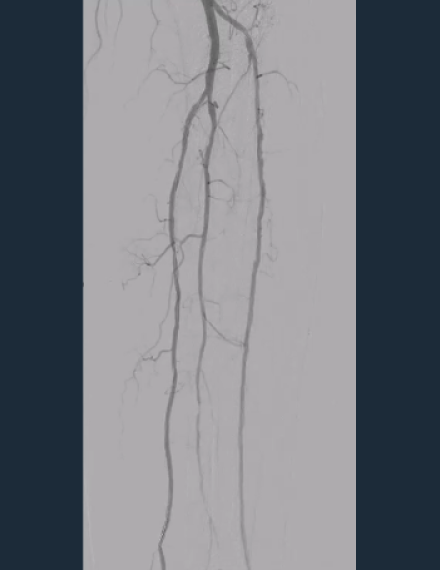
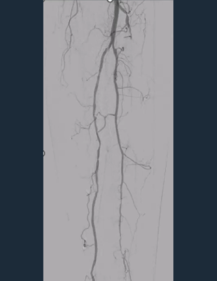
This case, courtesy of Dr. Maria Antonella Ruffino, Interventional Radiology, Lugano, features an ​83-year-old male patient with type 2 diabetes, dyslipidemia, and hypertension. He is suffering from Rutherford class 5 peripheral artery disease (PAD) and is presenting a heel ulcer.

  • Pre-procedure
    Baseline angio shows a total occlusion of the anterior tibial artery (ATA), with a diffuse lesion of 30 cm length.​ Ankle-brachial index (ABI) pre procedure: 0.30.
  • Pre-procedure
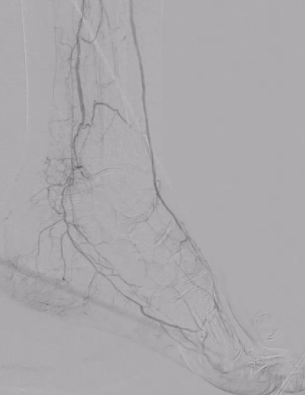
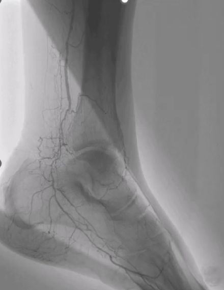
    Image shows poor distal run-off at the dorsalis pedis artery. ​
  • Shockwave Javelin Treatment
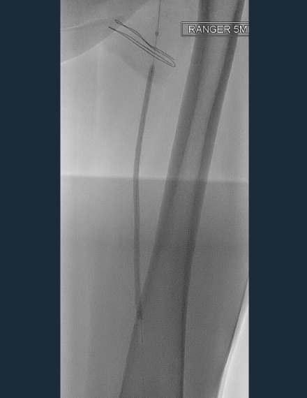
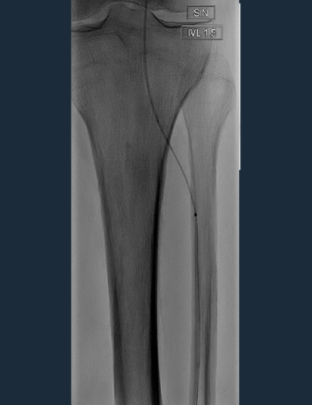
    After crossing with a 0.014 guidewire, 2.0 mm percutaneous transluminal angioplasty (PTA) balloon couldn’t cross at the origin of the ATA. ​Shockwave Javelin Peripheral was used to modify the calcium and cross the occlusion. ​
  • Shockwave Javelin Treatment
    120 pulses were delivered along the proximal and medial segment of the ATA. ​Shockwave Javelin Peripheral allowed to modify calcium along the lesion, enabling effective treatment with 2.5 mm PTA balloon.
  • Post-procedure
    Final angio shows restored flow of the anterior tibial artery. ABI post procedure: 0.75.
  • Post-procedure
    Foot angio shows direct flow from the anterior tibial artery into the dorsalis pedis and increased perfusion to the heel.

Case courtesy of Dr. Maria Antonella Ruffino, Interventional Radiology, Lugano.

Dr. Maria Antonella Ruffino is a paid consultant of Shockwave Medical.

This case, courtesy of Dr. Ashish Patel from St Thomas’, London, features a 71-year-old female patient, a smoker with hypertension Rutherford category 5 peripheral artery disease (PAD). The patient presents left hallux gangrene and severe rest pain and had no previous treatments. ​

  • Pre-procedure
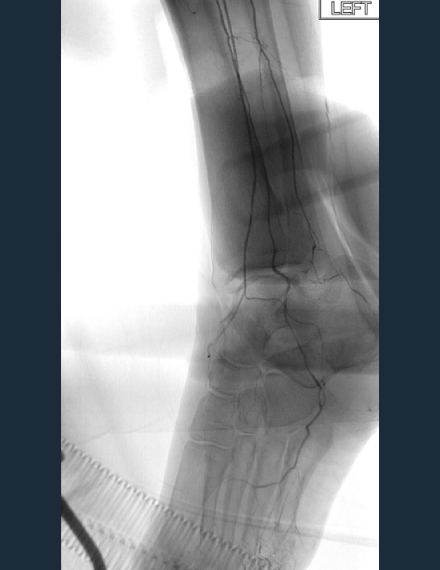
    Baseline angio shows occluded lateral plantar and dorsalis pedis arteries. ​Ankle-brachial index (ABI) pre-procedure: 0.4.
  • DCB Treatment
    Superficial femoral artery (SFA) and popliteal artery presenting some fibrotic plaque were treated with 5.0 mm plain old balloon angioplasty (POBA) and drug-coated balloon (DCB) to improve flow.
  • Shockwave Javelin Treatment
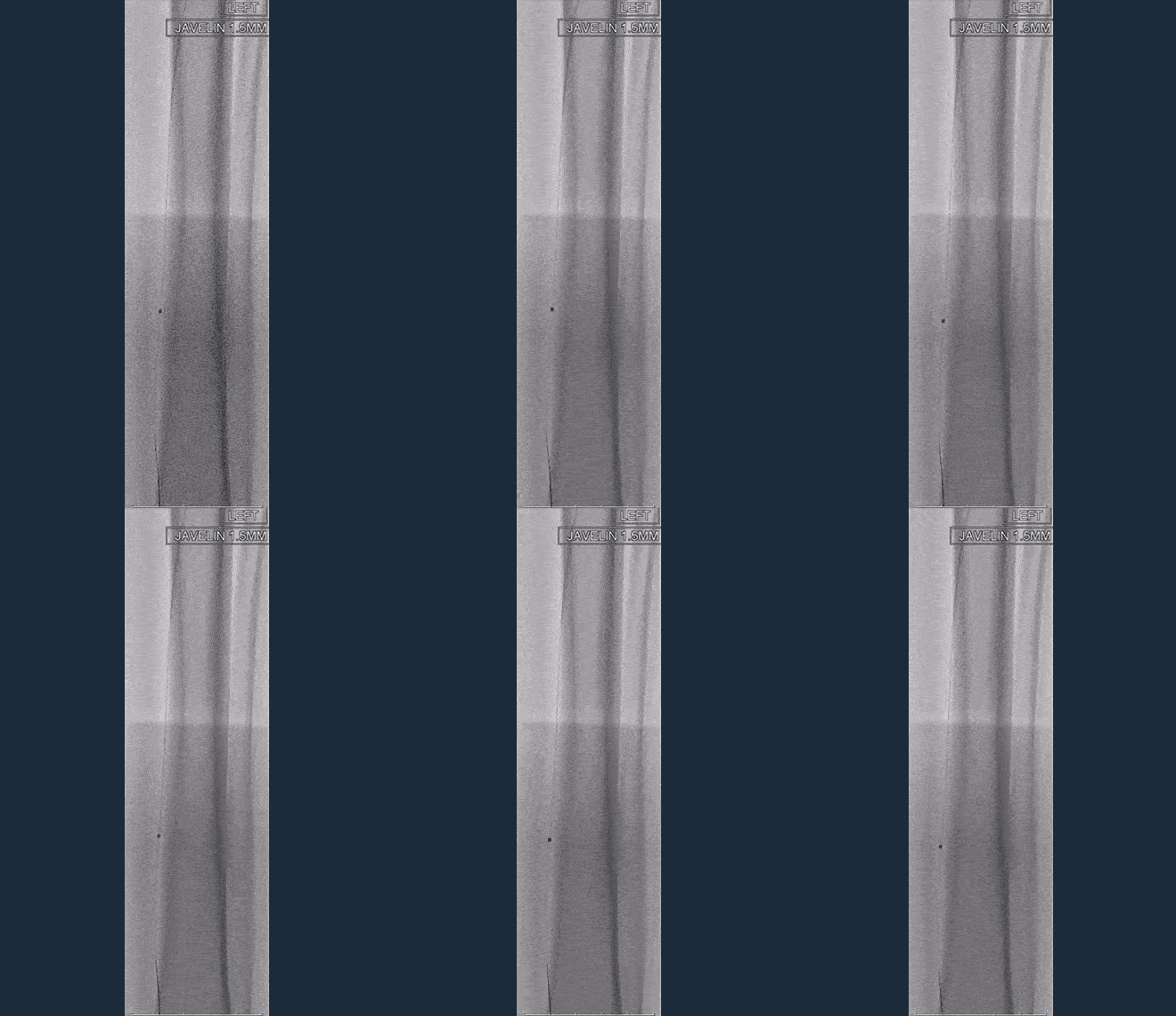
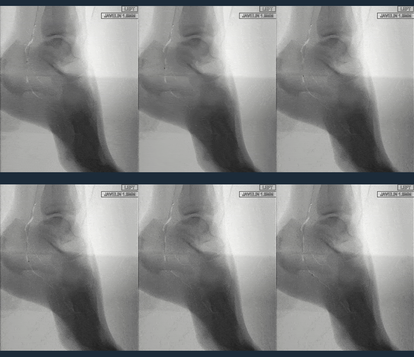
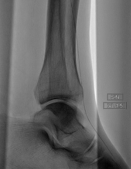
    The lesion was wire crossable but not device crossable (IVUS did not cross).​ Shockwave Javelin Peripheral was able to advance through the lesion.
  • Shockwave Javelin Treatment
    Shockwave Javelin Peripheral was advanced via the posterior tibial artery and through the common plantar artery.
  • Shockwave Javelin Treatment
    120 pulses of Shockwave Javelin Peripheral delivered across the occluded lateral plantar and dorsalis pedis arteries.
  • Final Treatment
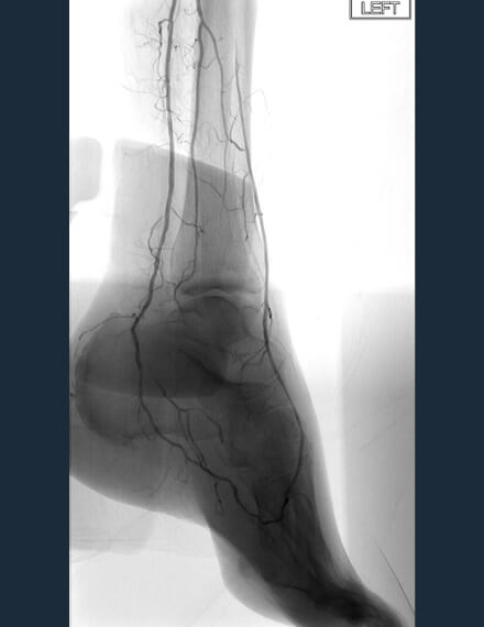
    After final treatment with 2.0 mm PTA balloon, final angio shows reconstitution of flow around the foot arch.​ ABI post-procedure: 0.8.

Case courtesy of Dr. Ashish Patel, St Thomas’, London.​

Dr. Ashish Patel is a paid consultant of Shockwave Medical.