Transfemoral access represents the gold standard for TAVI, with TF access showing a significant reduction in complications and mortality in comparison to alternative access strategies, which add additional complexity and risk to the procedure. Despite the wide acceptance that the transfemoral route provides the safest access for patients, the approach can be hindered by calcified iliac arteries inhibiting delivery of large bore devices by restricting the luminal diameter and reducing vessel compliance. If calcium is not treated adequately prior to TAVI delivery, this can lead to complications such as flow limiting dissection, vessel rupture and unsuccessful delivery of the implant.

Watch as Dr. Ole De Backer of Rigshospitalet, Copenhagen walks you through how intravascular lithotripsy (IVL) has expanded the boundaries of transfemoral TAVI. During this tutorial series Dr. De Backer will provide a detailed explanation of why, how and when IVL can be utilised to improve procedural safety and outcomes for patients undergoing transfemoral TAVI where calcium proves to be an inhibiting factor.

Why Take the Transfemoral Route? IVL Pre TAVI Tutorial Chapter 1

Unique Mechanism of Action to Treat Calcium: IVL Pre TAVI Tutorial Chapter 2

The Copenhagen Experience: IVL Pre TAVI Tutorial Chapter 3

How to Use IVL to Facilitate TF Access: IVL Pre TAVI Tutorial Chapter 4

IVL in Practice, Patient Selection and Cases: IVL Pre TAVI Tutorial Chapter 5

In addition to the tutorial series, check out the recent publication from Frontiers of Cardiovascular Medicine, which provides a comprehensive overview from Copenhagen of how IVL is simplifying extremely challenging transfemoral access in TAVI: https://doi.org/10.3389/fcvm.2021.739750


The physicians featured are paid consultants for Shockwave Medical.

Important Safety Information

Please contact your local Shockwave representative for specific country availability and refer to the Shockwave S4, Shockwave M5 and Shockwave M5+ instructions for use containing important safety information.

Caution: Federal law (USA) restricts this device to sale by or on the order of a physician.

Indication for Use – The Shockwave Medical Intravascular Lithotripsy (IVL) System is intended for lithotripsy-enhanced balloon dilatation of lesions, including calcified lesions, in the peripheral vasculature, including the iliac, femoral, ilio-femoral, popliteal, infra-popliteal, and renal arteries. Not for use in the coronary or cerebral vasculature.

Contraindications – Do not use if unable to pass 0.014 guidewire across the lesion • Not intended for treatment of in-stent restenosis or in coronary, carotid, or cerebrovascular arteries.

Warnings – Only to be used by physicians who are familiar with interventional vascular procedures • Physicians must be trained prior to use of the device • Use the Generator in accordance with recommended settings as stated in the Operator’s Manual.

Precautions – Use only the recommended balloon inflation medium • Appropriate anticoagulant therapy should be administered by the physician • Decision regarding use of distal protection should be made based on physician assessment of treatment lesion morphology.

Adverse Effects – Possible adverse effects consistent with standard angioplasty include: • Access site complications • Allergy to contrast or blood thinners • Arterial bypass surgery • Bleeding complications • Death • Fracture of guidewire or device • Hypertension/Hypotension • Infection/sepsis • Placement of a stent • Renal failure • Shock/pulmonary edema • Target vessel stenosis or occlusion • Vascular complications. Risks unique to the device and its use: • Allergy to catheter material(s) • Device malfunction or failure • Excess heat at target site.

Prior to use, please reference the Instructions for Use for more information on indications, contraindications, warnings, precautions, and adverse events. www.shockwavemedical.com

Calcium Masterclass Class 6 featuring James Spratt, Margaret McEntegart, Philippe Genereux, Michael Haude & Holger Nef.


Shockwave C2 and Shockwave C2+ Safety Information

In the United States: Rx only

Indications for Use— The Shockwave Intravascular Lithotripsy (IVL) System with the Shockwave C2 and C2+ Coronary IVL Catheter is indicated for lithotripsy-enabled, low-pressure balloon dilatation of severely calcified, stenotic de novo coronary arteries prior to stenting.

Contraindications— The Shockwave C2 and C2+ Coronary IVL System is contraindicated for the following: This device is not intended for stent delivery. This device is not intended for use in carotid or cerebrovascular arteries.

Warnings— Use the IVL Generator in accordance with recommended settings as stated in the Operator’s Manual. The risk of a dissection or perforation is increased in severely calcified lesions undergoing percutaneous treatment, including IVL. Appropriate provisional interventions should be readily available. Balloon loss of pressure was associated with a numerical increase in dissection which was not statistically significant and was not associated with MACE. Analysis indicates calcium length is a predictor of dissection and balloon loss of pressure. IVL generates mechanical pulses which may cause atrial or ventricular capture in bradycardic patients. In patients with implantable pacemakers and defibrillators, the asynchronous capture may interact with the sensing capabilities. Monitoring of the electrocardiographic rhythm and continuous arterial pressure during IVL treatment is required. In the event of clinically significant hemodynamic effects, temporarily cease delivery of IVL therapy.

Precautions— Only to be used by physicians trained in angiography and intravascular coronary procedures. Use only the recommended balloon inflation medium. Hydrophilic coating to be wet only with normal saline or water and care must be taken with sharp objects to avoid damage to the hydrophilic coating. Appropriate anticoagulant therapy should be administered by the physician. Precaution should be taken when treating patients with previous stenting within 5mm of target lesion.

Potential adverse effects consistent with standard based cardiac interventions include– Abrupt vessel closure – Allergic reaction to contrast medium, anticoagulant and/or antithrombotic therapy-Aneurysm-Arrhythmia-Arteriovenous fistula-Bleeding complications-Cardiac tamponade or pericardial effusion-Cardiopulmonary arrest-Cerebrovascular accident (CVA)-Coronary artery/vessel occlusion, perforation, rupture or dissection-Coronary artery spasm-Death-Emboli (air, tissue, thrombus or atherosclerotic emboli)-Emergency or nonemergency coronary artery bypass surgery-Emergency or nonemergency percutaneous coronary intervention-Entry site complications-Fracture of the guide wire or failure/malfunction of any component of the device that may or may not lead to device embolism, dissection, serious injury or surgical intervention-Hematoma at the vascular access site(s)- Hemorrhage-Hypertension/Hypotension-Infection/sepsis/fever-Myocardial Infarction-Myocardial Ischemia or unstable angina-Pain-Peripheral Ischemia-Pseudoaneurysm-Renal failure/insufficiency-Restenosis of the treated coronary artery leading to revascularization-Shock/pulmonary edema-Slow flow, no reflow, or abrupt closure of coronary artery-Stroke Thrombus-Vessel closure, abrupt-Vessel injury requiring surgical repair-Vessel dissection, perforation, rupture, or spasm.

Risks identified as related to the device and its use: Allergic/immunologic reaction to the catheter material(s) or coating-Device malfunction, failure, or balloon loss of pressure leading to device embolism, dissection, serious injury or surgical intervention-Atrial or ventricular extrasystole-Atrial or ventricular capture.

Prior to use, please reference the Instructions for Use for more information on indications, contraindications, warnings, precautions and adverse events. www.shockwavemedical.com/IFU.

Dr. Srinivasa Potluri, Chair of the Department of Cardiology and Director of the Cardiac Catheterization Laboratory at Baylor Scott and White, The Heart Hospital in Plano, speaks about his valuable experience with Shockwave coronary IVL. Dr. Potluri also shares a clinical case with severe ostial RCA and the value of coronary IVL in vessel preparation strategy in the presence of a heavy coronary calcium burden.

Download the PDF or read The Calcium Corner online here.

This article was developed in collaboration with Cath Lab Digest to bring you the latest calcium insights from the U.S. coronary interventional experts who know it best. A new webpage on the CLD website, The Calcium Corner, contains a series of articles where operators share their perspective on the challenges and treatment of coronary artery calcium. Read about experts’ clinical experience, case studies and treatment algorithms with Shockwave coronary IVL – hope you enjoy the content.


Dr. Srinivasa Potluri is a paid consultant for Shockwave Medical.

Coronary Important Safety Information:

In the United States: Rx only.

Indications for Use—The Shockwave Intravascular Lithotripsy (IVL) System with the Shockwave C2 Coronary IVL Catheter is indicated for lithotripsy-enabled, low-pressure balloon dilatation of severely calcified, stenotic de novo coronary arteries prior to stenting.

Contraindications—The Shockwave C2 Coronary IVL System is contraindicated for the following: This device is not intended for stent delivery. This device is not intended for use in carotid or cerebrovascular arteries.

Warnings— Use the IVL Generator in accordance with recommended settings as stated in the Operator’s Manual. The risk of a dissection or perforation is increased in severely calcified lesions undergoing percutaneous treatment, including IVL. Appropriate provisional interventions should be readily available. Balloon loss of pressure was associated with a numerical increase in dissection which was not statistically significant and was not associated with MACE.  Analysis indicates calcium length is a predictor of dissection and balloon loss of pressure.  IVL generates mechanical pulses which may cause atrial or ventricular capture in bradycardic patients. In patients with implantable pacemakers and defibrillators, the asynchronous capture may interact with the sensing capabilities. Monitoring of the electrocardiographic rhythm and continuous arterial pressure during IVL treatment is required.  In the event of clinically significant hemodynamic effects, temporarily cease delivery of IVL therapy.

Precautions— Only to be used by physicians trained in angiography and intravascular coronary procedures. Use only the recommended balloon inflation medium. Hydrophilic coating to be wet only with normal saline or water and care must be taken with sharp objects to avoid damage to the hydrophilic coating. Appropriate anticoagulant therapy should be administered by the physician. Precaution should be taken when treating patients with previous stenting within 5mm of target lesion.

Potential adverse effects consistent with standard based cardiac interventions include– Abrupt vessel closure – Allergic reaction to contrast medium, anticoagulant and/or antithrombotic therapy-Aneurysm-Arrhythmia-Arteriovenous fistula-Bleeding complications-Cardiac tamponade or pericardial effusion-Cardiopulmonary arrest-Cerebrovascular accident (CVA)-Coronary artery/vessel occlusion, perforation, rupture or dissection-Coronary artery spasm-Death-Emboli (air, tissue, thrombus or atherosclerotic emboli)-Emergency or non-emergency coronary artery bypass surgery-Emergency or non-emergency percutaneous coronary intervention-Entry site complications-Fracture of the guide wire or failure/malfunction of any component of the device that may or may not lead to device embolism, dissection, serious injury or surgical intervention-Hematoma at the vascular access site(s)-Hemorrhage-Hypertension/Hypotension-Infection/sepsis/fever-Myocardial Infarction-Myocardial Ischemia or unstable angina-Pain-Peripheral Ischemia-Pseudoaneurysm-Renal failure/insufficiency-Restenosis of the treated coronary artery leading to revascularization-Shock/pulmonary edema-Slow flow, no reflow, or abrupt closure of coronary artery-Stroke-Thrombus-Vessel closure, abrupt-Vessel injury requiring surgical repair-Vessel dissection, perforation, rupture, or spasm. Risks identified as related to the device and its use: Allergic/immunologic reaction to the catheter material(s) or coating-Device malfunction, failure, or balloon loss of pressure leading to device embolism, dissection, serious injury or surgical intervention-Atrial or ventricular extrasystole-Atrial or ventricular capture.

Prior to use, please reference the Instructions for Use for more information on warnings, precautions and adverse events.  https://shockwavemedical.com/IFU

Please contact your local Shockwave representative for specific country availability and refer to the Shockwave C2 instructions for use containing important safety information.

Q&A with Dr. Ziad Ali about the DISRUPT CAD Pooled OCT Eccentric vs Concentric Analysis at TCT21.

Following its presentation at TCT21 in a moderated poster session, we caught up with Ziad Ali, M.D., DPhil, Director of the DeMatteis Cardiovascular Institute and Investigational Interventional Cardiology at St Francis Hospital & Heart Center, to get his thoughts on the implications of the concentric vs eccentric calcium analysis among those patients enrolled in the DISRUPT CAD clinical program.

Why is eccentric calcium so challenging to modify?

Dr. Ali: Simple. It’s hard to modify. Balloon based modification leads to the creation of a dissection at the site of minimal resistance. That is the fibro-calcific interface, the place where the fibrous tissue meets the calcium. This dissection allows vessel expansion, but the calcium remains unmodified. After stenting the calcium protrudes back into towards the lumen, reducing the potential MSA. With atherectomy, the wire and thus burr must be biased into the calcium. If it is not, there is no chance of lesion modification. With IVL, most of the energy is lost into the soft tissue. So if there is only 90 degrees of calcification, i.e 25% of the vessel circumference, 75% of the energy is dissipated. More specifically, the maximum energy in IVL is immediately perpendicular to the electrode.

Why is the DISRUPT CAD OCT Pooled analysis important for current clinical practice?

Dr. Ali: Simple. There is power in numbers. These data show categorically that the predominant mechanism of IVL is calcium fracture, and that the more the calcium the greater the fracture. The ability of IVL to create luminal gain in severe calcification is unquestionable. Overall the mean stent expansion and stent expansion at the site of max calcium was greater than 100%. The fact that the minimal lumen area was never at the site of maximum calcification is proof in and of itself.

What were the findings in the OCT analysis in concentric and eccentric lesions? How do you explain these results?

Dr. Ali: Fractures are much less common in eccentric rather than concentric lesions. But that’s ok, because you need less fractures in eccentric calcium because the rest of the artery is conformable. In general, lesion modification for an eccentric calcified lesion is unnecessary, unless it’s a calcified nodule. That being said, within a heavily calcified segment of vessel, there will be multiple morphologies along the length of that lesion. Some concentric, some eccentric. IVL helps all the way along the length by creating lots of fractures at the more concentric sites and less fractures, because less are needed, at the eccentric sites. The take home message is that even in eccentric lesions there was a consistent improvement in stent expansion and luminal gain compared to concentric lesions. So IVL liberates vascular compliance through calcium fracture appropriate to need.

From your perspective, how do these outcomes compare to what you see in the clinic?

Dr. Ali: We are definitely still learning, but the clinical experience is very similar to the trial results. Not surprising, there was tremendous consistency in terms of safety and efficacy among all of DISRUPT CAD studies so we wouldn’t expect that different in the real world. At St Francis we have done over 100 commercial cases and one of our more noticeable findings is how well tolerated IVL is in the very high-risk patient. Very low EF, high EDP, severe PAH, multivessel disease. We don’t see the same hemodynamic fluctuations we see sometimes with other lesion prep strategies.

Are there any coronary IVL best practices that will help interventionalists achieve similar results to what was reported?

Dr. Ali: Image image image. Intravascular imaging determines the severity and distribution of calcium. Using the St Francis Calcium Scores can help determine whether or not you even need advanced lesion prep. By OCT, if the calcium is 5 mm long, 0.5 mm thick and 50% of the arc, advanced prep is necessary. By IVUS, circumferential, 270 degrees for 5 mm, < 3.5 mm at the site of calcification and calcified nodule, advanced prep is necessary. Remember the more the rock the better the shock!


Dr. Ali is a paid consultant for Shockwave Medical.

Important Safety Information

Please contact your local Shockwave representative for specific country availability and refer to the Shockwave C2 instructions for use containing important safety information.

Rx only

Indications for Use—The Shockwave Intravascular Lithotripsy (IVL) System with the Shockwave C2 Coronary IVL Catheter is indicated for lithotripsy-enabled, low-pressure balloon dilatation of severely calcified, stenotic de novo coronary arteries prior to stenting.

Contraindications—The Shockwave C2 Coronary IVL System is contraindicated for the following: This device is not intended for stent delivery. This device is not intended for use in carotid or cerebrovascular arteries.

Warnings— Use the IVL Generator in accordance with recommended settings as stated in the Operator’s Manual. The risk of a dissection or perforation is increased in severely calcified lesions undergoing percutaneous treatment, including IVL. Appropriate provisional interventions should be readily available. Balloon loss of pressure was associated with a numerical increase in dissection which was not statistically significant and was not associated with MACE.  Analysis indicates calcium length is a predictor of dissection and balloon loss of pressure.  IVL generates mechanical pulses which may cause atrial or ventricular capture in bradycardic patients. In patients with implantable pacemakers and defibrillators, the asynchronous capture may interact with the sensing capabilities. Monitoring of the electrocardiographic rhythm and continuous arterial pressure during IVL treatment is required.  In the event of clinically significant hemodynamic effects, temporarily cease delivery of IVL therapy.

Precautions— Only to be used by physicians trained in angiography and intravascular coronary procedures. Use only the recommended balloon inflation medium. Hydrophilic coating to be wet only with normal saline or water and care must be taken with sharp objects to avoid damage to the hydrophilic coating. Appropriate anticoagulant therapy should be administered by the physician. Precaution should be taken when treating patients with previous stenting within 5mm of target lesion.

Potential adverse effects consistent with standard based cardiac interventions include– Abrupt vessel closure – Allergic reaction to contrast medium, anticoagulant and/or antithrombotic therapy-Aneurysm-Arrhythmia-Arteriovenous fistula-Bleeding complications-Cardiac tamponade or pericardial effusion-Cardiopulmonary arrest-Cerebrovascular accident (CVA)-Coronary artery/vessel occlusion, perforation, rupture or dissection-Coronary artery spasm-Death-Emboli (air, tissue, thrombus or atherosclerotic emboli)-Emergency or non-emergency coronary artery bypass surgery-Emergency or non-emergency percutaneous coronary intervention-Entry site complications-Fracture of the guide wire or failure/malfunction of any component of the device that may or may not lead to device embolism, dissection, serious injury or surgical intervention-Hematoma at the vascular access site(s)-Hemorrhage-Hypertension/Hypotension-Infection/sepsis/fever-Myocardial Infarction-Myocardial Ischemia or unstable angina-Pain-Peripheral Ischemia-Pseudoaneurysm-Renal failure/insufficiency-Restenosis of the treated coronary artery leading to revascularization-Shock/pulmonary edema-Slow flow, no reflow, or abrupt closure of coronary artery-Stroke-Thrombus-Vessel closure, abrupt-Vessel injury requiring surgical repair-Vessel dissection, perforation, rupture, or spasm.

Risks identified as related to the device and its use: Allergic/immunologic reaction to the catheter material(s) or coating-Device malfunction, failure, or balloon loss of pressure leading to device embolism, dissection, serious injury or surgical intervention-Atrial or ventricular extrasystole-Atrial or ventricular capture.

Prior to use, please reference the Instructions for Use for more information on warnings, precautions and adverse events.  https://shockwavemedical.com/IFU

Q&A with Dr. Akiko Maehara about the DISRUPT CAD Pooled OCT Calcific Nodules Analysis at TCT21


How do you define a calcific nodule and what makes them so difficult to modify?

Dr. Maehara: As defined by OCT, the eruptive calcific nodule is accumulation of small calcium fragments underlying the calcified plaque typically protruding the lumen. Nodular calcium1 (healed calcified nodule, i.e., calcified nodule with thick fibrous cap) is probably more difficult to modify because calcium is hard structured and because nodules protrude into the lumen which makes fracturing them very difficult with sub-optimal stent expansion.


Why is the DISRUPT CAD OCT analysis of calcified nodules relevant to today’s practice?

Dr. Maehara: This analysis is relevant because lesions with calcified nodules present with poor long-term outcomes, even with a good minimum stent area (MSA) at index procedure. There is a very robust recent Japanese articlepublished in Atherosclerosis by H. Sugane et al which shows that when we compare severe calcified lesions with or without calcified nodules, the lesions with calcified nodules have poor outcomes compared with the lesions without calcified nodules. While MSA is the most important factor to predict the future event, long-term outcomes are still poor in the presence of calcified nodules regardless of the index MSA. In another Japanese article published in Atherosclerosis by H. Sugane et al using the ISR captured by directional coronary atherectomy3, when they studied the ISR cases, the lesions with calcified nodules showed them protruding through the stent struts, and the calcified nodule was pushed out. This is not something we have seen before, and we are still learning about it.


From your perspective, what did we learn from this study on the impact of coronary IVL on nodular calcium?

Dr. Maehara: The nodular analysis is still a preliminary finding as it is looking at procedural results, so we should acknowledge that we need longer-term clinical follow-up of these patients. That said, my sense looking at this OCT analysis and considering my experience with nodular lesions treated without IVL, is that IVL is disrupting the calcified fragments that are bonded together to create the calcific nodule. IVL seems to be reshaping the calcified nodule which might mean more symmetric stent expansion and less protruding calcium through the struts during follow-up, which theoretically may lead to better long-term outcomes than treatments with other calcium modification therapies. We really have to see clinically good outcomes in the IVL cohort compared to a non-IVL cohort or possibly look into the OCT again in the chronic stage to see if the nodules are protruding inside the stent. That’s kind of the data that is needed to support this type of hypothesis, which will come with longer follow-up.


How would you explain IVL’s ability to successfully modify nodules in this analysis to the interventional cardiologist who do not consider the technology a great tool for this calcium morphology?

Dr. Maehara: We all know these cohorts have very poor outcomes even in comparison to severely calcified lesions without nodules. While the calcified nodule looks like a calcium rock, pathologically, the very dense calcium is small calcium fragments connected with fibrous tissue. As such, the IVL acoustic energy can disrupt the small fragments affecting the calcified nodule. Balloons, on the other hand, are subject to wire bias and are not changing the shape of the calcified nodule. Ablation therapies are polishing only one side of the nodule, just 15% of it.4 To be able to ablate more of the nodule with atherectomy, say 50%, you would have to ablate multiple times which we can’t typically do. This is why IVL disrupts calcified nodules much easier than other calcium modification modalities.


Given that these are acute procedural outcomes, what is important to look at next from a research perspective with coronary IVL in calcific nodules? 

Dr. Maehara: We have to see clinical outcomes in the long-term. No matter how good stent expansion is, these lesions have poor outcomes because of protruding nodules. Will fracturing the calcified nodule help the long-term outcome? Will the acute appearance correlate with the outcomes? We will see over time.

In addition, we need to better understand different types of calcified nodules and what are the optimal outcomes for each type of nodule, and which calcium treatment device might be ideal for each type.

It is also important to study patients having hemodialysis as they are more likely to have accumulation of calcification, and as a result, more calcified nodules. They are truly a unique cohort with very poor outcomes. This is even more critical to look into for international regions where transplants are not an option.


1: Torii et al, Pathology of Calcified Nodule J A C C V O L . 7 7 , N O . 1 3 , 2 0 2 1 A P R I L 6 , 2 0 2 1 : 1 5 9 9 – 6 1 1
2: H. Sugane et al, Atherosclerosis, https://doi.org/10.1016/j.atherosclerosis.2020.11.005
3: Nakamura et al, J Am Heart Assoc. 2020;9:e016595. DOI: 10.1161/JAHA.120.016595 1
4: Yamamoto et al, Catheter Cardiovasc Interv. 2018;1–8. 

Dr. Maehara is a paid consultant for Shockwave Medical.


Coronary Important Safety Information:

In the United States: Rx only.

Indications for Use—The Shockwave Intravascular Lithotripsy (IVL) System with the Shockwave C2 Coronary IVL Catheter is indicated for lithotripsy-enabled, low-pressure balloon dilatation of severely calcified, stenotic de novo coronary arteries prior to stenting.

Contraindications—The Shockwave C2 Coronary IVL System is contraindicated for the following: This device is not intended for stent delivery. This device is not intended for use in carotid or cerebrovascular arteries.

Warnings— Use the IVL Generator in accordance with recommended settings as stated in the Operator’s Manual. The risk of a dissection or perforation is increased in severely calcified lesions undergoing percutaneous treatment, including IVL. Appropriate provisional interventions should be readily available. Balloon loss of pressure was associated with a numerical increase in dissection which was not statistically significant and was not associated with MACE.  Analysis indicates calcium length is a predictor of dissection and balloon loss of pressure.  IVL generates mechanical pulses which may cause atrial or ventricular capture in bradycardic patients. In patients with implantable pacemakers and defibrillators, the asynchronous capture may interact with the sensing capabilities. Monitoring of the electrocardiographic rhythm and continuous arterial pressure during IVL treatment is required.  In the event of clinically significant hemodynamic effects, temporarily cease delivery of IVL therapy.

Precautions— Only to be used by physicians trained in angiography and intravascular coronary procedures. Use only the recommended balloon inflation medium. Hydrophilic coating to be wet only with normal saline or water and care must be taken with sharp objects to avoid damage to the hydrophilic coating. Appropriate anticoagulant therapy should be administered by the physician. Precaution should be taken when treating patients with previous stenting within 5mm of target lesion.

Potential adverse effects consistent with standard based cardiac interventions include– Abrupt vessel closure – Allergic reaction to contrast medium, anticoagulant and/or antithrombotic therapy-Aneurysm-Arrhythmia-Arteriovenous fistula-Bleeding complications-Cardiac tamponade or pericardial effusion-Cardiopulmonary arrest-Cerebrovascular accident (CVA)-Coronary artery/vessel occlusion, perforation, rupture or dissection-Coronary artery spasm-Death-Emboli (air, tissue, thrombus or atherosclerotic emboli)-Emergency or non-emergency coronary artery bypass surgery-Emergency or non-emergency percutaneous coronary intervention-Entry site complications-Fracture of the guide wire or failure/malfunction of any component of the device that may or may not lead to device embolism, dissection, serious injury or surgical intervention-Hematoma at the vascular access site(s)-Hemorrhage-Hypertension/Hypotension-Infection/sepsis/fever-Myocardial Infarction-Myocardial Ischemia or unstable angina-Pain-Peripheral Ischemia-Pseudoaneurysm-Renal failure/insufficiency-Restenosis of the treated coronary artery leading to revascularization-Shock/pulmonary edema-Slow flow, no reflow, or abrupt closure of coronary artery-Stroke-Thrombus-Vessel closure, abrupt-Vessel injury requiring surgical repair-Vessel dissection, perforation, rupture, or spasm. Risks identified as related to the device and its use: Allergic/immunologic reaction to the catheter material(s) or coating-Device malfunction, failure, or balloon loss of pressure leading to device embolism, dissection, serious injury or surgical intervention-Atrial or ventricular extrasystole-Atrial or ventricular capture.

Prior to use, please reference the Instructions for Use for more information on warnings, precautions and adverse events.  https://shockwavemedical.com/IFU

Please contact your local Shockwave representative for specific country availability and refer to the Shockwave C2 instructions for use containing important safety information.


Q&A with Dr. Dean Kereiakes about the DISRUPT CAD III 1yr Data at TCT21

Following its presentation at TCT21 in a featured clinical research session, we caught up with DISRUPT CAD III co-principal investigator, Dean Kereiakes, M.D., FACC, FSCAI, Medical Director of The Christ Hospital Heart and Vascular Center and the Christ Hospital Research Institute; Professor of Clinical Medicine, The Ohio State University, to get his thoughts on the data and what stands out from his perspective since the primary 30-day outcomes were presented at TCT20.


What is the significance of the one-year follow-up data from the DISRUPT CAD III study, which was included by TCT as one of the meeting’s best abstracts in the featured clinical research session?

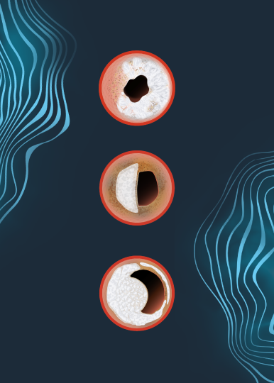
Dr. Kereiakes: I think it’s very significant that it shows sustained and persistent relative benefit of IVL for lesion preparation prior to coronary stenting. Since this is the first robust one-year data that has been presented on coronary IVL, I think that’s very important as there were concerns, as there have been with the use of ablative technologies, that in late follow-up there might be some loss of the benefit that might have been predicted by the MSA and percent stent expansion at the initial procedure. These were phenomenal levels from the OCT sub study – 102% stent expansion and MSA of 6.5mm2 at the site of maximum lesion calcification. The fact that we were able to achieve these excellent levels, one would predict based on all other previous imaging studies, that there should be a low rate of TLR and stent thrombosis, which is exactly what we found.


Were there any other concerns that you had about the potential one-year outcomes going into TCT?

Dr. Kereiakes: Well, some had expressed concerns that there might be a negative impact of the energy generated by IVL like we see with the friction induced by rotational and orbital atherectomy. The reassuring answer is it doesn’t look like it as the data are great. When, at 1-year, you have an ID-TLR of 4.3% and a total stent thrombosis rate of 1.1% with only one patient (0.3%) having a stent thrombosis beyond 30 days, that bodes very well.


Was there anything surprising to you about the data itself?

Dr. Kereiakes: No, I actually thought it was predictable based on the optimized stent implantation results we were able to achieve by pretreating these severely calcified lesions – 100% of which were classified as severely calcified as adjudicated by an independent core lab.


Now that durable one-year outcomes have been shown, where do we go from here from a clinical research perspective?

Dr. Kereiakes: I think the next step is to analyze the post-market approval study being done through the ACC CathPCI registry, which is very innovative. I think this is going to be very helpful to expand the observations using IVL for lesion preparation in a much broader population of real-world patients.


Given that this was a single arm study, how do you contextualize the results of the one-year findings?

Dr. Kereiakes: You know I think without a randomized comparator it’s always challenging and difficult. Recall that we set up the best performance goals you could possibly set up using a similar population, similar definitions and similar endpoints, to be identical to ORBIT II which was the pivotal trial for FDA approval of orbital atherectomy. This was probably the best designed non-randomized comparator possible. That said, although it remains a cross trial, non-randomized comparison, when you look at the one year data, you’ve got Disrupt CAD III at 13.8% MACE rate and 16.9% for ORBIT II. This is a very intriguing, hypothesis-generating but non-randomized comparison.


For those physicians that may have been waiting for longer term data to prove out the effectiveness of the therapy, what would you tell them now that the one-year data is available?

Dr. Kereiakes: I’d tell them to look at the data and look at the best data from the other sources of calcium modifying technologies – take in the totality of the data. That means peri-procedural results to 30-days and at one year. There’s never been a cohort of patients with more severely calcified target lesions than those enrolled in DISRUPT CAD III for U.S. FDA approval of coronary IVL. When you look at the substrate that was enrolled and you look at the peri-procedural outcomes, with zero perforations, zero abrupt closures, zero no re-flow with IVL alone and only one patient with an Ellis type 1 micro-perforation following stent deployment which sealed by itself out of the entire series. When I look at these data I say you have a low complication rate, great outcomes at 30 days that now persist out to one year. Knowing these data, going forward I would hesitate to approach severely calcified target lesions without IVL.


Is there anything else that stood out to you about the IVL data presented at TCT?

Dr. Kereiakes The other thing that I would honestly say, that’s really hard to recreate, is that when you look at all of the IVL data, we have 47 sites in four countries with DISRUPT CAD III and an additional pooled analysis of data from 72 sites in 12 countries. When you look at all of these different sites across multiple continents and countries, there is remarkable consistency in the safety and effectiveness endpoints achieved. I think that reflects the ease-of-use of IVL. It’s a balloon. Every interventionalist, I don’t care what subspecialty you are, uses balloons. And that’s the relative safety of the technology as well. Delivering high technology in a primitive delivery system. It doesn’t matter whether you’re in Europe or U.S. Doesn’t matter if you’re in a big center, or little center. The beauty of this observation is the consistency of safety and effectiveness. We found no differences with IVL. I call IVL the great equalizer.


Dr. Dean Kereiakes is a paid consultant for Shockwave Medical.

Coronary Important Safety Information:

In the United States: Rx only.

Indications for Use—The Shockwave Intravascular Lithotripsy (IVL) System with the Shockwave C2 Coronary IVL Catheter is indicated for lithotripsy-enabled, low-pressure balloon dilatation of severely calcified, stenotic de novo coronary arteries prior to stenting.

Contraindications—The Shockwave C2 Coronary IVL System is contraindicated for the following: This device is not intended for stent delivery. This device is not intended for use in carotid or cerebrovascular arteries.

Warnings— Use the IVL Generator in accordance with recommended settings as stated in the Operator’s Manual. The risk of a dissection or perforation is increased in severely calcified lesions undergoing percutaneous treatment, including IVL. Appropriate provisional interventions should be readily available. Balloon loss of pressure was associated with a numerical increase in dissection which was not statistically significant and was not associated with MACE.  Analysis indicates calcium length is a predictor of dissection and balloon loss of pressure.  IVL generates mechanical pulses which may cause atrial or ventricular capture in bradycardic patients. In patients with implantable pacemakers and defibrillators, the asynchronous capture may interact with the sensing capabilities. Monitoring of the electrocardiographic rhythm and continuous arterial pressure during IVL treatment is required.  In the event of clinically significant hemodynamic effects, temporarily cease delivery of IVL therapy.

Precautions— Only to be used by physicians trained in angiography and intravascular coronary procedures. Use only the recommended balloon inflation medium. Hydrophilic coating to be wet only with normal saline or water and care must be taken with sharp objects to avoid damage to the hydrophilic coating. Appropriate anticoagulant therapy should be administered by the physician. Precaution should be taken when treating patients with previous stenting within 5mm of target lesion.

Potential adverse effects consistent with standard based cardiac interventions include– Abrupt vessel closure – Allergic reaction to contrast medium, anticoagulant and/or antithrombotic therapy-Aneurysm-Arrhythmia-Arteriovenous fistula-Bleeding complications-Cardiac tamponade or pericardial effusion-Cardiopulmonary arrest-Cerebrovascular accident (CVA)-Coronary artery/vessel occlusion, perforation, rupture or dissection-Coronary artery spasm-Death-Emboli (air, tissue, thrombus or atherosclerotic emboli)-Emergency or non-emergency coronary artery bypass surgery-Emergency or non-emergency percutaneous coronary intervention-Entry site complications-Fracture of the guide wire or failure/malfunction of any component of the device that may or may not lead to device embolism, dissection, serious injury or surgical intervention-Hematoma at the vascular access site(s)-Hemorrhage-Hypertension/Hypotension-Infection/sepsis/fever-Myocardial Infarction-Myocardial Ischemia or unstable angina-Pain-Peripheral Ischemia-Pseudoaneurysm-Renal failure/insufficiency-Restenosis of the treated coronary artery leading to revascularization-Shock/pulmonary edema-Slow flow, no reflow, or abrupt closure of coronary artery-Stroke-Thrombus-Vessel closure, abrupt-Vessel injury requiring surgical repair-Vessel dissection, perforation, rupture, or spasm. Risks identified as related to the device and its use: Allergic/immunologic reaction to the catheter material(s) or coating-Device malfunction, failure, or balloon loss of pressure leading to device embolism, dissection, serious injury or surgical intervention-Atrial or ventricular extrasystole-Atrial or ventricular capture.

Prior to use, please reference the Instructions for Use for more information on warnings, precautions and adverse events.  https://shockwavemedical.com/IFU

Please contact your local Shockwave representative for specific country availability and refer to the Shockwave C2 instructions for use containing important safety information.


Q&A with Dr. Yasin Hussain about the DISRUPT CAD Pooled Female vs. Male Analysis at TCT21


What do we know about gender analyses from large PCI studies previously conducted?

Dr. Hussain: In general, moderately to severely calcified lesions are associated with higher rates of target lesion failure, myocardial infarction, stent thrombosis, and cardiac death both in hospital and long-term and independently predict ischemia-driven revascularization in both men and women. Typically, women undergoing PCI have more comorbidities and worse periprocedural complications. When it comes to PCI of severely calcified lesions, women have as much as a two-fold higher risk of coronary perforations and a five-fold higher risk of tamponade compared with men, and the risk of in-hospital mortality is reported to be 20% higher compared to men.


From your perspective, what were the key takeaways from this analysis?

Dr. Hussain: This pooled analysis of the DISRUPT CAD trials is the largest to evaluate the use of Shockwave IVL to treat severely calcified coronary lesions. The key findings were: first: IVL is safe and effective in both men and women; second: IVL treatment of severe calcification resulted in the lowest observed rates of procedural complications in both men and women compared to other atheroablative options.


What is the impact of the Gender Analysis of the DISRUPT CAD Pooled Studies on current practice in both men and women patient populations?

Dr. Hussain: IVL appears to be the great equalizer of outcomes between men and women showing for the first time lower procedural complications in women compared to men. IVL should be considered first line treatment for severely calcified lesions especially in women given their increased risk of severe procedural complications and the poor associated clinical outcomes including heightened mortality risk.


How do outcomes from DISRUPT CAD Pooled Gender Analysis compare to previous gender studies conducted with calcium modification tools?

Dr. Hussain: Several small studies have looked at the differences in outcomes using rotational atherectomy and orbital atherectomy based on sex. Atheroablative devices have two-to-five-fold higher periprocedural complications in women compared with men. IVL in our study shows low and similar results in men and women. However, it is important to highlight that this observation is based on small, non-randomized series and will require additional confirmation.


What should interventional cardiologists expect to learn next about Shockwave coronary IVL in females – where should the research take us?

Dr. Hussain: Future studies should compare the safety and efficacy in a randomized manner of an IVL versus a standard atheroablative treatment strategy to confirm and understand the generalizability of our preliminary results.


Dr. Yasin Hussain is a paid consultant for Shockwave Medical.

Coronary Important Safety Information:

In the United States: Rx only.

Indications for Use—The Shockwave Intravascular Lithotripsy (IVL) System with the Shockwave C2 Coronary IVL Catheter is indicated for lithotripsy-enabled, low-pressure balloon dilatation of severely calcified, stenotic de novo coronary arteries prior to stenting.

Contraindications—The Shockwave C2 Coronary IVL System is contraindicated for the following: This device is not intended for stent delivery. This device is not intended for use in carotid or cerebrovascular arteries.

Warnings— Use the IVL Generator in accordance with recommended settings as stated in the Operator’s Manual. The risk of a dissection or perforation is increased in severely calcified lesions undergoing percutaneous treatment, including IVL. Appropriate provisional interventions should be readily available. Balloon loss of pressure was associated with a numerical increase in dissection which was not statistically significant and was not associated with MACE.  Analysis indicates calcium length is a predictor of dissection and balloon loss of pressure.  IVL generates mechanical pulses which may cause atrial or ventricular capture in bradycardic patients. In patients with implantable pacemakers and defibrillators, the asynchronous capture may interact with the sensing capabilities. Monitoring of the electrocardiographic rhythm and continuous arterial pressure during IVL treatment is required.  In the event of clinically significant hemodynamic effects, temporarily cease delivery of IVL therapy.

Precautions— Only to be used by physicians trained in angiography and intravascular coronary procedures. Use only the recommended balloon inflation medium. Hydrophilic coating to be wet only with normal saline or water and care must be taken with sharp objects to avoid damage to the hydrophilic coating. Appropriate anticoagulant therapy should be administered by the physician. Precaution should be taken when treating patients with previous stenting within 5mm of target lesion.

Potential adverse effects consistent with standard based cardiac interventions include– Abrupt vessel closure – Allergic reaction to contrast medium, anticoagulant and/or antithrombotic therapy-Aneurysm-Arrhythmia-Arteriovenous fistula-Bleeding complications-Cardiac tamponade or pericardial effusion-Cardiopulmonary arrest-Cerebrovascular accident (CVA)-Coronary artery/vessel occlusion, perforation, rupture or dissection-Coronary artery spasm-Death-Emboli (air, tissue, thrombus or atherosclerotic emboli)-Emergency or non-emergency coronary artery bypass surgery-Emergency or non-emergency percutaneous coronary intervention-Entry site complications-Fracture of the guide wire or failure/malfunction of any component of the device that may or may not lead to device embolism, dissection, serious injury or surgical intervention-Hematoma at the vascular access site(s)-Hemorrhage-Hypertension/Hypotension-Infection/sepsis/fever-Myocardial Infarction-Myocardial Ischemia or unstable angina-Pain-Peripheral Ischemia-Pseudoaneurysm-Renal failure/insufficiency-Restenosis of the treated coronary artery leading to revascularization-Shock/pulmonary edema-Slow flow, no reflow, or abrupt closure of coronary artery-Stroke-Thrombus-Vessel closure, abrupt-Vessel injury requiring surgical repair-Vessel dissection, perforation, rupture, or spasm. Risks identified as related to the device and its use: Allergic/immunologic reaction to the catheter material(s) or coating-Device malfunction, failure, or balloon loss of pressure leading to device embolism, dissection, serious injury or surgical intervention-Atrial or ventricular extrasystole-Atrial or ventricular capture.

Prior to use, please reference the Instructions for Use for more information on warnings, precautions and adverse events.  https://shockwavemedical.com/IFU

Please contact your local Shockwave representative for specific country availability and refer to the Shockwave C2 instructions for use containing important safety information.

In the recent Cardiovascular Revascularization Medicine article, “First United States experience with RotaShock: A case series,” Drs. Gautam Kumar and Rajesh Sachdeva of Emory and Atlanta VA Medical Center recount three successful cases using the RotaShock technique, a combination of rotational atherectomy with IVL for severely calcified vessels. Following its publication, we engaged them in a highly educational Q&A discussion about how the two technologies are very complementary to each other, and the new strategy can be used either electively or as a rescue. We hope you enjoy their additional perspective.


Out of all of your IVL cases performed, what percentage of your cases have been Rota-Shock and do you expect this rate to decrease or increase over time?

Drs. Kumar & Sachdeva: We have done rotational atherectomy plus IVL in about 10% of our cases. We have several other modalities available in our cath lab and we are also actively participating in randomized trials for calcified coronary lesions.


The complementary relationship between atherectomy and IVL that you reference in your manuscript – is it equally effective with both rotational and orbital atherectomy, or is one preferred?

Drs. Kumar & Sachdeva: Rotational atherectomy (RA) works predominantly against superficial calcium but orbital atherectomy does have an effect on fragmenting the deep calcium in addition to sanding the superficial calcium. This has been described in at least a few studies that have done intravascular imaging in conjunction with orbital atherectomy like in our own series – Desai R et al. Plaque modification of severely calcified coronary lesions via orbital atherectomy: Single-center observations from a complex Veterans Affairs cohort. Health Sci Rep. 2018 Oct 27;1(12):e99. The complementary relationship between RA and IVL seems to make intuitive sense.


You make a keen distinction in your study between “Elective” and “Rescue” RotaShock approaches – what percentage of your RotaShock cases would you expect to see in category?

Drs. Kumar & Sachdeva: Having several years of experience treating calcified lesions in the VA population, we have quickly learnt that planning a combination approach upfront is a better methodology especially when you have information from intravascular imaging or CCTA. There are unusual circumstances where IVL will be used as a rescue therapy and we envision that with increased usage of pre-PCI imaging, this will be necessary less often.


In choosing the “Elective” RotaShock strategy, what are you looking for on angio or intravascular imaging to help make that decision?

Drs. Kumar & Sachdeva: The most important factor that we use to identify an elective Rota-Shock case on angiography is the size of the vessel. In general, for a large left main coronary artery (5-6 mm diameter) or a proximal left anterior descending artery (4-5 mm diameter), it is going to be hard to assume that plaque modification with a 1.5 mm burr alone will be adequate prior to stenting to maximize MSA. The depth of calcification is an important factor is determining whether plaque fracturing is adequate, and OCT is superior to IVUS for the assessment of this as the acoustic shadowing of the proximal edge of the calcium precludes depth assessment with IVUS. Post-atherectomy, we can assess the extent of plaque fracturing with OCT or with IVUS as well.


In the “Rescue” RotaShock cases, what were your previous treatment option before IVL was available?

Drs. Kumar & Sachdeva: One could consider rotational atherectomy with a larger size burr prior to stenting. Sometimes, this may be difficult in radial cases as a 7 Fr guide catheter may not have been used initially and thus burr size would have been limited to 1.5 mm as an example. If orbital atherectomy had been chosen, we could consider going back and doing more runs and consider high speed runs at 120k rpm if the vessel was large. Prior to IVL, most operators would have used specialty balloons with variable results. Some operators may consider laser depending upon availability.


Are there any tips or tricks that you use to optimize outcomes in your RotaShock cases?

Drs. Kumar & Sachdeva: Mandatory use of imaging – either IVUS or preferably, OCT is essential. We generally try to evaluate each case pre-plaque modification (if we are able to cross the lesion), post-plaque modification (to assess the adequacy of calcium fracturing prior to stenting) and then post stenting (to assess stent expansion and apposition).


As you’ve gained more experience with RotaShock, what do you know now that you wish you would have known before your first case?

Drs. Kumar & Sachdeva: We are still early in our experience with this technique and continue to learn from each case.


Dr. Gautam Kumar and Dr. Rajesh Sachdeva are paid consultants for Shockwave Medical.

Coronary Important Safety Information:

In the United States: Rx only.

Indications for Use—The Shockwave Intravascular Lithotripsy (IVL) System with the Shockwave C2 Coronary IVL Catheter is indicated for lithotripsy-enabled, low-pressure balloon dilatation of severely calcified, stenotic de novo coronary arteries prior to stenting.

Contraindications—The Shockwave C2 Coronary IVL System is contraindicated for the following: This device is not intended for stent delivery. This device is not intended for use in carotid or cerebrovascular arteries.

Warnings— Use the IVL Generator in accordance with recommended settings as stated in the Operator’s Manual. The risk of a dissection or perforation is increased in severely calcified lesions undergoing percutaneous treatment, including IVL. Appropriate provisional interventions should be readily available. Balloon loss of pressure was associated with a numerical increase in dissection which was not statistically significant and was not associated with MACE.  Analysis indicates calcium length is a predictor of dissection and balloon loss of pressure.  IVL generates mechanical pulses which may cause atrial or ventricular capture in bradycardic patients. In patients with implantable pacemakers and defibrillators, the asynchronous capture may interact with the sensing capabilities. Monitoring of the electrocardiographic rhythm and continuous arterial pressure during IVL treatment is required.  In the event of clinically significant hemodynamic effects, temporarily cease delivery of IVL therapy.

Precautions— Only to be used by physicians trained in angiography and intravascular coronary procedures. Use only the recommended balloon inflation medium. Hydrophilic coating to be wet only with normal saline or water and care must be taken with sharp objects to avoid damage to the hydrophilic coating. Appropriate anticoagulant therapy should be administered by the physician. Precaution should be taken when treating patients with previous stenting within 5mm of target lesion.

Potential adverse effects consistent with standard based cardiac interventions include– Abrupt vessel closure – Allergic reaction to contrast medium, anticoagulant and/or antithrombotic therapy-Aneurysm-Arrhythmia-Arteriovenous fistula-Bleeding complications-Cardiac tamponade or pericardial effusion-Cardiopulmonary arrest-Cerebrovascular accident (CVA)-Coronary artery/vessel occlusion, perforation, rupture or dissection-Coronary artery spasm-Death-Emboli (air, tissue, thrombus or atherosclerotic emboli)-Emergency or non-emergency coronary artery bypass surgery-Emergency or non-emergency percutaneous coronary intervention-Entry site complications-Fracture of the guide wire or failure/malfunction of any component of the device that may or may not lead to device embolism, dissection, serious injury or surgical intervention-Hematoma at the vascular access site(s)-Hemorrhage-Hypertension/Hypotension-Infection/sepsis/fever-Myocardial Infarction-Myocardial Ischemia or unstable angina-Pain-Peripheral Ischemia-Pseudoaneurysm-Renal failure/insufficiency-Restenosis of the treated coronary artery leading to revascularization-Shock/pulmonary edema-Slow flow, no reflow, or abrupt closure of coronary artery-Stroke-Thrombus-Vessel closure, abrupt-Vessel injury requiring surgical repair-Vessel dissection, perforation, rupture, or spasm. Risks identified as related to the device and its use: Allergic/immunologic reaction to the catheter material(s) or coating-Device malfunction, failure, or balloon loss of pressure leading to device embolism, dissection, serious injury or surgical intervention-Atrial or ventricular extrasystole-Atrial or ventricular capture.

Prior to use, please reference the Instructions for Use for more information on warnings, precautions and adverse events.  https://shockwavemedical.com/IFU

Please contact your local Shockwave representative for specific country availability and refer to the Shockwave C2 instructions for use containing important safety information.

Calcium causes a magnitude of problems in EVAR and TEVAR procedures. Calcium can inhibit stent graft delivery leading to significant dissection or perforation of the iliac arteries. Calcium can also affect expansion of iliac limbs leading to increased risk of under expansion and stent graft thrombosis.

Watch as Dr. Stefano Fazzini, Tor Vergata University Hospital, Italy walks you through how Shockwave IVL can help alleviate access challenges while minimizing complications and the need for secondary interventions.


Dr. Stefano Fazzini is a paid consultant for Shockwave Medical.

Important Safety Information

Caution: Federal law (USA) restricts this device to sale by or on the order of a physician.

Indication for Use – The Shockwave Medical Intravascular Lithotripsy (IVL) System is intended for lithotripsy-enhanced balloon dilatation of lesions, including calcified lesions, in the peripheral vasculature, including the iliac, femoral, ilio-femoral, popliteal, infra-popliteal, and renal arteries.  Not for use in the coronary or cerebral vasculature.

Contraindications – Do not use if unable to pass 0.014 guidewire across the lesion • Not intended for treatment of in-stent restenosis or in coronary, carotid, or cerebrovascular arteries.

Warnings – Only to be used by physicians who are familiar with interventional vascular procedures • Physicians must be trained prior to use of the device • Use the Generator in accordance with recommended settings as stated in the Operator’s Manual

Precautions – Use only the recommended balloon inflation medium • Appropriate anticoagulant therapy should be administered by the physician • Decision regarding use of distal protection should be made based on physician assessment of treatment lesion morphology

Adverse Effects – Possible adverse effects consistent with standard angioplasty include: • Access site complications • Allergy to contrast or blood thinners • Arterial bypass surgery • Bleeding complications • Death • Fracture of guidewire or device • Hypertension/Hypotension • Infection/sepsis • Placement of a stent • Renal failure • Shock/pulmonary edema • Target vessel stenosis or occlusion • Vascular complications.

Risks unique to the device and its use: • Allergy to catheter material(s) • Device malfunction or failure • Excess heat at target site

Prior to use, please reference the Instructions for Use for more information on indications, contraindications, warnings, precautions, and adverse events. www.shockwavemedical.com

Calcium Masterclass Class 8 featuring Ashok Seth, Benjamin Honton, James Spratt & Jonathan Hill.


The physicians featured are paid consultants for Shockwave Medical.