Recently, Dr. James Spratt, consultant cardiologist at St George’s University NHS Trust in London and founder of Optima Education, and other leading UK cardiologists published their experience with Intravascular Lithotripsy (IVL) in the left main in EuroIntervention: Intravascular lithotripsy for the treatment of calcific distal left main disease.

We had the privilege of connecting with Dr. Spratt to learn more about his publication and get some of his key takeaways as one of the most prolific IVL users since the introduction of the technology nearly two years ago:

Question: What concerns or considerations do you have about modifying calcium in the left main?

Dr. Spratt: The limitations in percutaneous treatment of the left main nearly always come down to the ability to achieve a certain size or volume in three key areas; one being the ostium of the LAD; second being the ostium of the circumflex; and the third area being called the polygon of confluence, which is basically where the LAD and the circumflex join into the distal end of the left main.

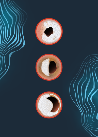
Normally, plaque is relatively compliant, so it can be shifted out of the way or stretched with balloon treatment. Calcium, however, is not compliant, and therefore not possible to shift it out of the way with just balloon treatment.

Prior to IVL, the only real significant tool we had to modify calcium was rotational atherectomy. The limitation with rotational atherectomy is first of all, the procedure in the left main can be especially hazardous because of the risk of distal no-reflow, but also, rotablation can’t work on lesions that it can’t ablate or touch. Therefore, if the lesion is not tight enough so that the burr doesn’t touch the lesion, then it doesn’t ablate it.

So prior to the development of IVL, while we could identify calcium, there was very little that we could do to modify it, particularly if it was deep-wall calcium.

Dr Spratt for Catalyst

Question: How does calcium most often present itself in the left main from your experience? Is it more proximal? More distal? Looking at that area in confluence, is it usually concentric or eccentric?

Dr. Spratt: Calcium is much more common in the bifurcation than the LMS shaft or ostium. Most of that is due to flow dynamics. Again, if you look at the bifurcation itself, the disease is more likely to be not at the carina, essentially the middle of the trousers of the two arteries, but at the sides. So as general rule, disease is more common at the bifurcation ~ 80% of total disease.

In terms of drilling down to the real granularity of looking at eccentricity versus concentricity, there really isn’t a lot of data to guide the answer. To date, it’s been primarily driven by anecdotal experience.

Question: What were your study’s goals – what did you seek to understand going into the research?

Dr. Spratt: The main goal of the study was to do two things, one to demonstrate feasibility in treating highly calcific lesions in patients who often didn’t have an alternative revascularization strategy and in whom previously only medical therapy was an option. And we knew that in that group, if they’re not treated with revascularization, they actually do very badly. So our goal was to demonstrate that they could be safely and effectively treated.

We understand the limitations of reporting a technique without any randomization, but nevertheless, given the novel nature of the technology and the importance of the topic, we felt that we could add value by reporting a relatively large number of patients with good results in 100% in patients of which, I would say, estimated 70 or 80% we would have no alternative form of treatment just one year ago.

Question: What were your study’s findings?

Dr. Spratt: The first thing to say is that we have very good surrogate markers of what constitutes the success for percutaneous revascularisation in the left main stem (LMS), and it’s not just looking at an angiogram and saying that looks good or that doesn’t look good. Unfortunately patient level experience isn’t granular enough to tell you how durable the result is likely to be. So, in other words, you can’t ask a patient how they feel the day after procedure and expect that to be a good guide of how they’ll feel in three or six months or a year or five years.

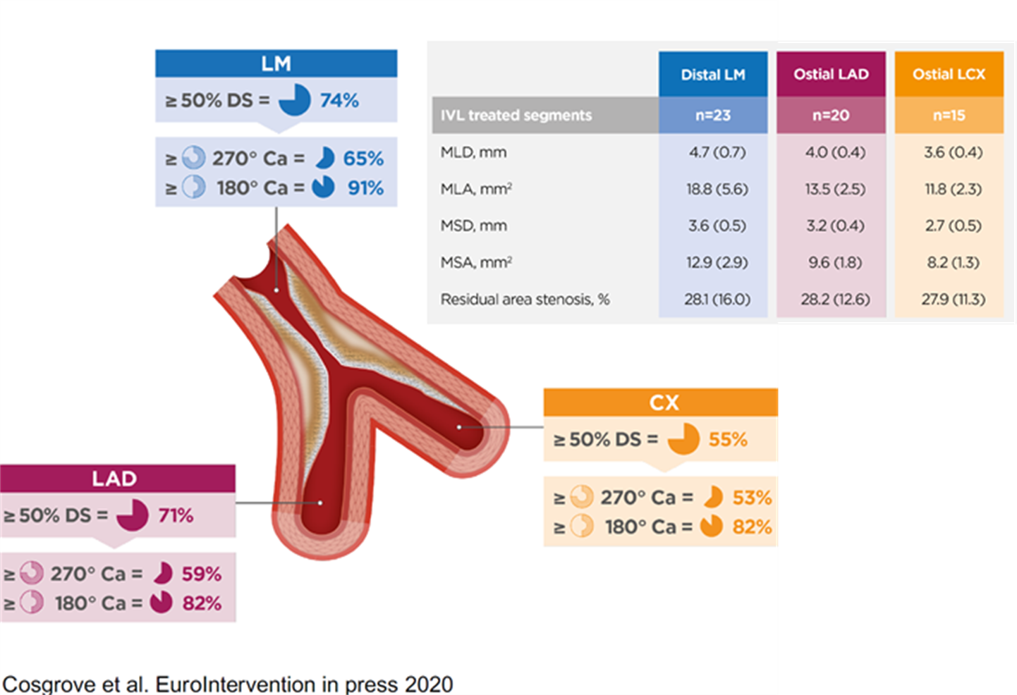
However measurements at the end of the procedure can be extrapolated to outcomes as far out as five years from now. Those measurements are very simply volume measurements or at least area measurements at the three critical areas: the LAD ostium, the circumflex ostium, and that polygon of confluence where the LM stem meets the LAD and circ. So one of our major goals was to show that in very severe calcific disease we could get areas there which we could extrapolate and predict a good three to five-year outcome for these patients.

And actually, even within our relatively small study, despite the fact that we were treating very severe disease, the average numbers we got in these areas with lithotripsy were actually higher than in both the two main left main stem papers published recently, NOBLE and EXCEL. So that was a major finding. (See graphic below for detailed area measurements.)

And the second major finding is that we could do that safely. There had been some considerations raised about the safety of prolonged balloon inflation in the left main stem that’s a necessary part of lithotripsy treatment. But we were fine, we were able to deliver lithotripsy without any complications whatsoever. And in the vast majority of our patients, they were able to be discharged the same day. And all of the patients have kept well since.

Dr James Spratt Graphic for Catalyst Post

Question: What further research needs remain regarding IVL in the LM?

Dr. Spratt: When introducing new technology, the first thing to demonstrate is safety. I think there’s an increasing body of evidence that lithotripsy is safe, but while safety is good and important, it’s not enough on its own. The second step is efficacy. You want to be sure that it’s efficacious. So I think within a very small number of patients, it was safe and efficacious. But for it to become standard of care, it’s going to need more evidence than we have currently.

So at present there’s good evidence that if you don’t have an alternative good revascularization strategy in somebody with highly calcific left main disease, and that alternative would be bypass surgery, then this works very well and it’s safe and it’s relatively straightforward to use.

And I think we must all be very careful and not too overreaching in what we claim from what is a relatively small study. I think it’s a positive result. It’s given us encouragement to push forward. And I think we will be looking to follow it up with a more robust reflection of what lithotripsy can deliver in this environment.

Question: Based on your personal use, are there any best practices that you’ve identified when using IVL in the left main that leads to a more efficacious result?

Dr. Spratt: Within my practice, which consists mostly of surgical turndowns, CTOs, and post-bypass CTOs, it’s very straightforward technology to use. I think it’s best complimented by an appreciation of where best to apply it. I think that’s best achieved by intravascular imaging.

And again, it’s best applied where the goal is fairly clear. And I think the left main is one of the clearest areas where if you achieve certain imaging defined goals, you can be very confident in what you tell your patient about the success of the procedure.

My advice to fellow interventionists would be carefully assess where you’re going to need IVL. You don’t want to use it in every patient. After you have used it, carefully assess the cracked calcium by intravascular imaging. Finally, once the stent has been implanted, carefully assess that you’ve achieved the goals that you set yourself out at the start, that you’ve got those three key measurements that you were aiming for. And then once you’ve done all that, then you can tell the patient with confidence that they should keep well for a long period of time.

Question: How do you size your IVL catheter in the LM, especially when you have a larger LM?

Dr. Spratt: As you will be aware the maximum size of IVL balloon is 4mm and the recommended sizing is 1:1 – it is extremely rare that this will fail to appose in the body of the LMS (even in a very large vessel due to the fact that the lesion is quite stenosed) & will be appropriately sized in the daughter vessels. If the vessel is above 5.0mm, one might consider using a 1:1 RVD-sized NC balloon after IVL to further open up the vessel after the calcium has been fractured to maximize lumen gain before stent implantation.


Important Safety Information – Coronary

Caution: In the United States, Shockwave C2 Coronary IVL catheters are investigational devices, limited by United States law to investigational use. DISRUPT CAD III Study

Shockwave C2 Coronary IVL catheters are commercially available in certain countries outside the U.S. Please contact your local Shockwave representative for specific country availability. The Shockwave C2 Coronary IVL catheters are indicated for lithotripsy-enhanced, low-pressure balloon dilatation of calcified, stenotic de novo coronary arteries prior to stenting. For the full IFU containing important safety information please visit: https://shockwavemedical.com/clinicians/international/coronary/shockwave-c2/

The story of how Shockwave intravascular lithotripsy (IVL) came into being is a paragon of true innovation – it could be a chapter out of Steven Johnson’s book, Where Good Ideas Come From. The founders demonstrate a textbook example of how the amalgamation of building liquid networks, developing the slow hunch, error, serendipity and expanding into the adjacent possible led to the disruptive innovation that is IVL.

See what role a child’s piece of chalk, organic chicken eggs and cadaver legs all play in Shockwave IVL’s journey from idea to reality in this fun video.


Important Safety Information

Caution: Federal law (USA) restricts this device to sale by or on the order of a physician.

Indication for Use – The Shockwave Medical Intravascular Lithotripsy (IVL) System is intended for lithotripsy-enhanced balloon dilatation of lesions, including calcified lesions, in the peripheral vasculature, including the iliac, femoral, ilio-femoral, popliteal, infra-popliteal, and renal arteries. Not for use in the coronary or cerebral vasculature.

Contraindications – Do not use if unable to pass 0.014 guidewire across the lesion • Not intended for treatment of in-stent restenosis or in coronary, carotid, or cerebrovascular arteries.

Warnings – Only to be used by physicians who are familiar with interventional vascular procedures • Physicians must be trained prior to use of the device • Use the Generator in accordance with recommended settings as stated in the Operator’s Manual

Precautions – Use only the recommended balloon inflation medium • Appropriate anticoagulant therapy should be administered by the physician • Decision regarding use of distal protection should be made based on physician assessment of treatment lesion morphology

Adverse Effects – Possible adverse effects consistent with standard angioplasty include: • Access site complications • Allergy to contrast or blood thinners • Arterial bypass surgery • Bleeding complications • Death • Fracture of guidewire or device • Hypertension/Hypotension • Infection/sepsis • Placement of a stent • Renal failure • Shock/pulmonary edema • Target vessel stenosis or occlusion • Vascular complications. Risks unique to the device and its use: • Allergy to catheter material(s) • Device malfunction or failure • Excess heat at target site

Prior to use, please reference the Instructions for Use for more information on indications, contraindications, warnings, precautions, and adverse events.

42 patients, 42 valves safely implanted through calcified iliofemoral access!

Read about the multicenter registry results of using Shockwave Intravascular Lithotripsy to preserve Transfemoral TAVR in a recent publication in JACC: Cardiovascular Interventions.

Read the publication here.


Important Safety Information

Caution: Federal law (USA) restricts this device to sale by or on the order of a physician. Indication for Use – The Shockwave Medical Intravascular Lithotripsy (IVL) System is intended for lithotripsy-enhanced balloon dilatation of lesions, including calcified lesions, in the peripheral vasculature, including the iliac, femoral, ilio-femoral, popliteal, infra-popliteal, and renal arteries.  Not for use in the coronary or cerebral vasculature.

Contraindications – Do not use if unable to pass 0.014 guidewire across the lesion • Not intended for treatment of in-stent restenosis or in coronary, carotid, or cerebrovascular arteries.

Warnings – Only to be used by physicians who are familiar with interventional vascular procedures • Physicians must be trained prior to use of the device • Use the Generator in accordance with recommended settings as stated in the Operator’s Manual

Precautions – Use only the recommended balloon inflation medium • Appropriate anticoagulant therapy should be administered by the physician • Decision regarding use of distal protection should be made based on physician assessment of treatment lesion morphology

Adverse Effects – Possible adverse effects consistent with standard angioplasty include: • Access site complications • Allergy to contrast or blood thinners • Arterial bypass surgery • Bleeding complications • Death • Fracture of guidewire or device • Hypertension/Hypotension • Infection/sepsis • Placement of a stent • Renal failure • Shock/pulmonary edema • Target vessel stenosis or occlusion • Vascular complications. Risks unique to the device and its use: • Allergy to catheter material(s) • Device malfunction or failure • Excess heat at target site

Prior to use, please reference the Instructions for Use for more information on indications, contraindications, warnings, precautions, and adverse events. www.shockwavemedical.com

The complete DISRUPT CAD II study findings have been published by the AHA’s journal, Circulation: Cardiovascular Interventions, and while it’s definitely worth the full read, we know that sometimes there just aren’t enough hours in the day. For a quick synopsis, we took the liberty to summarize some of the key data points into a visual-friendly infographic for you to review some of the study’s most important findings.

Download the Infographic PDF Here

Infographic titled 'Disrupt CAD II: A New Wave of Data.'


Coronary Important Safety Information:

In the United States: Rx only.

Indications for Use—The Shockwave Intravascular Lithotripsy (IVL) System with the Shockwave C2 Coronary IVL Catheter is indicated for lithotripsy-enabled, low-pressure balloon dilatation of severely calcified, stenotic de novo coronary arteries prior to stenting.

Contraindications—The Shockwave C2 Coronary IVL System is contraindicated for the following: This device is not intended for stent delivery. This device is not intended for use in carotid or cerebrovascular arteries.

Warnings— Use the IVL Generator in accordance with recommended settings as stated in the Operator’s Manual. The risk of a dissection or perforation is increased in severely calcified lesions undergoing percutaneous treatment, including IVL. Appropriate provisional interventions should be readily available. Balloon loss of pressure was associated with a numerical increase in dissection which was not statistically significant and was not associated with MACE.  Analysis indicates calcium length is a predictor of dissection and balloon loss of pressure.  IVL generates mechanical pulses which may cause atrial or ventricular capture in bradycardic patients. In patients with implantable pacemakers and defibrillators, the asynchronous capture may interact with the sensing capabilities. Monitoring of the electrocardiographic rhythm and continuous arterial pressure during IVL treatment is required.  In the event of clinically significant hemodynamic effects, temporarily cease delivery of IVL therapy.

Precautions— Only to be used by physicians trained in angiography and intravascular coronary procedures. Use only the recommended balloon inflation medium. Hydrophilic coating to be wet only with normal saline or water and care must be taken with sharp objects to avoid damage to the hydrophilic coating. Appropriate anticoagulant therapy should be administered by the physician. Precaution should be taken when treating patients with previous stenting within 5mm of target lesion.

Potential adverse effects consistent with standard based cardiac interventions include– Abrupt vessel closure – Allergic reaction to contrast medium, anticoagulant and/or antithrombotic therapy-Aneurysm-Arrhythmia-Arteriovenous fistula-Bleeding complications-Cardiac tamponade or pericardial effusion-Cardiopulmonary arrest-Cerebrovascular accident (CVA)-Coronary artery/vessel occlusion, perforation, rupture or dissection-Coronary artery spasm-Death-Emboli (air, tissue, thrombus or atherosclerotic emboli)-Emergency or non-emergency coronary artery bypass surgery-Emergency or non-emergency percutaneous coronary intervention-Entry site complications-Fracture of the guide wire or failure/malfunction of any component of the device that may or may not lead to device embolism, dissection, serious injury or surgical intervention-Hematoma at the vascular access site(s)-Hemorrhage-Hypertension/Hypotension-Infection/sepsis/fever-Myocardial Infarction-Myocardial Ischemia or unstable angina-Pain-Peripheral Ischemia-Pseudoaneurysm-Renal failure/insufficiency-Restenosis of the treated coronary artery leading to revascularization-Shock/pulmonary edema-Slow flow, no reflow, or abrupt closure of coronary artery-Stroke-Thrombus-Vessel closure, abrupt-Vessel injury requiring surgical repair-Vessel dissection, perforation, rupture, or spasm. Risks identified as related to the device and its use: Allergic/immunologic reaction to the catheter material(s) or coating-Device malfunction, failure, or balloon loss of pressure leading to device embolism, dissection, serious injury or surgical intervention-Atrial or ventricular extrasystole-Atrial or ventricular capture.

Prior to use, please reference the Instructions for Use for more information on warnings, precautions and adverse events.  https://shockwavemedical.com/IFU

Please contact your local Shockwave representative for specific country availability and refer to the Shockwave C2 instructions for use containing important safety information.

Case submitted by Nelson Bernardo, MD, MedStar Health

Summary: Patient was considered high surgical risk due to severity of calcium and involvement of profunda. Shockwave IVL treatment of common femoral artery (CFA) and proximal superficial femoral artery (SFA), followed by treatment of profunda with the same device. Final result shows no dissection, perforation, emboli, need for additional devices and full resolution of symptoms.

Case submitted by Marianne Brodmann, MD, University of Graz

Summary: Proximal total occlusion and distal sub-total occlusion of popliteal artery. Severe calcification throughout. 4.5 mm x 60 mm Shockwave IVL delivered distally, followed by 5.5 mm x 60 mm IVL proximally. Final result shows <30% residual with IVL alone, no complications and no implants.

Case submitted by Sarang Mangalmurti, MD, Mainline Health

Summary: Severe concentric calcification of anterior tibial artery with 100% occlusion. 3.0 mm x 40 mm Shockwave IVL delivered into AT with no difficulty. >12 cm of AT treated. <20% residual stenosis followed stand-alone IVL with no complications.

Case submitted by Roberto Ferraresi, MD, Humanitas Gavazzeni

Summary: CLTI patient (Rutherford 5) with foot ulceration. Diagnostic angiogram shows near-occlusion of distal AT. 2.5 mm percutaneous transluminal angioplasty (PTA) of AT and dorsalis pedia attempted with near-immediate recoil and unsatisfactory result. 2.5 mm x 40 mm Shockwave IVL introduced into distal AT. Post-IVL angiogram shows resolution of stenosis with good distal flow into dorsalis pedis and no complications.


Case submitted by Jean Fajadet and Bruno Farah, Clinique Pasteur

Summary: Severely calcified distal left main and proximal left anterior descending (LAD) lesion; operators were hesitant to use rota; Shockwave IVL advanced with no pre-dilation required; 3.5 x12 mm IVL treatment performed and balloon waist resolved after 20 pulses; final result of widely patent artery with TIMI 3 flow post-stent (3.5 mm) with 4.5 mm NC post-dil.


Case submitted by Javier Escaned, San Carlos Hospital

Summary: Severely calcified mid-left anterior descending artery (LAD) involving septal branch; patient was high-bleeding risk and IABP used for hemodynamic support; operators wanted to avoid distal embolization for this patient and chose Shockwave IVL; 3.0 mm IVL catheter used across diffuse calcified disease; significant fractures seen under OCT in multiple planes; circumferential and well-apposed 3.0 x 32 mm drug-eluting stent (DES).