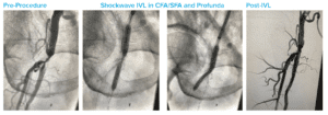
Case submitted by Marianne Brodmann, MD, University of Graz

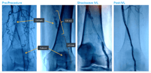
Summary: Proximal total occlusion and distal sub-total occlusion of popliteal artery. Severe calcification throughout. 4.5 mm x 60 mm Shockwave IVL delivered distally, followed by 5.5 mm x 60 mm IVL proximally. Final result shows <30% residual with IVL alone, no complications and no implants.

Case submitted by Sarang Mangalmurti, MD, Mainline Health

Summary: Severe concentric calcification of anterior tibial artery with 100% occlusion. 3.0 mm x 40 mm Shockwave IVL delivered into AT with no difficulty. >12 cm of AT treated. <20% residual stenosis followed stand-alone IVL with no complications.

Case submitted by Roberto Ferraresi, MD, Humanitas Gavazzeni

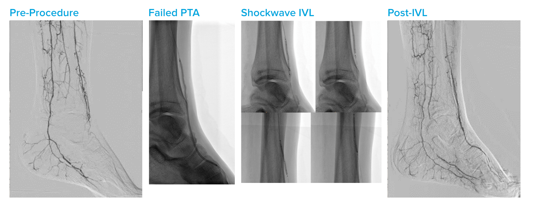
Summary: CLTI patient (Rutherford 5) with foot ulceration. Diagnostic angiogram shows near-occlusion of distal AT. 2.5 mm percutaneous transluminal angioplasty (PTA) of AT and dorsalis pedia attempted with near-immediate recoil and unsatisfactory result. 2.5 mm x 40 mm Shockwave IVL introduced into distal AT. Post-IVL angiogram shows resolution of stenosis with good distal flow into dorsalis pedis and no complications.


Case submitted by Jean Fajadet and Bruno Farah, Clinique Pasteur

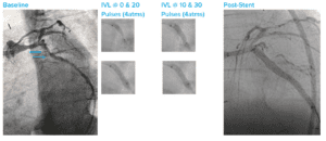
Summary: Severely calcified distal left main and proximal left anterior descending (LAD) lesion; operators were hesitant to use rota; Shockwave IVL advanced with no pre-dilation required; 3.5 x12 mm IVL treatment performed and balloon waist resolved after 20 pulses; final result of widely patent artery with TIMI 3 flow post-stent (3.5 mm) with 4.5 mm NC post-dil.


Case submitted by Javier Escaned, San Carlos Hospital

Summary: Severely calcified mid-left anterior descending artery (LAD) involving septal branch; patient was high-bleeding risk and IABP used for hemodynamic support; operators wanted to avoid distal embolization for this patient and chose Shockwave IVL; 3.0 mm IVL catheter used across diffuse calcified disease; significant fractures seen under OCT in multiple planes; circumferential and well-apposed 3.0 x 32 mm drug-eluting stent (DES).


Case submitted by Javier Escaned, San Carlos Hospital

Summary: Highly angulated left circumflex artery (LCX) with lesions proximal and distal; OCT wouldn’t cross either lesion; guideliner-assisted Shockwave IVL catheter delivery followed by successful IVL therapy; the case was completed with easy deployment of two long drug-eluting stents (DES).


Case submitted by Jonathan Hill, King’s College

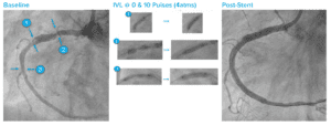
Summary: Multi-lesion right coronary artery (RCA); couldn’t advance guideliner to distal lesion despite predilation; advanced 3.5 mm Shockwave IVL catheter as far as possible (1); delivered one cycle (10 pulses) and vessel opened; pulled back to the ostium (2) and vessel opened after one cycle (10 pulses); advanced to distal lesion (3) and vessel opened after one cycle (10 pulses); easily delivered 80 mm of drug-eluting stent (DES).


Case submitted by Antonio Colombo, San Raffaele

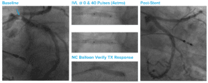
Summary: Large eccentric lesion at left circumflex (LCX) ostium; delivered four cycles with 3.5 mm Shockwave IVL catheter; full balloon expansion (and associated lower endoflator pressure) were noted; verified treatment response using a non-compliant balloon; then used the same IVL device to successfully treat the left main (LM).


Case submitted by Jonathan Hill, King’s College

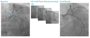
Summary: Unprotected left main (LM) trifurcation with heavy calcium throughout LM, left anterior descending artery (LAD), left circumflex artery (LCX), and at ostia of trifurcation; hemodynamic support with temporary pump; Shockwave IVL administered along LAD, LCX, LM and at the ostia of LCX, LAD, and Ramus; drug-eluting stents (DES) placed in LM and all three vessels with good expansion and apposition (demonstrated with OCT).


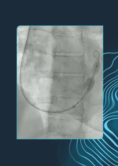
Case submitted by Brian Kolski, MD, St. Joseph Hospital

Summary: 85-year-old woman with critical aortic stenosis and severe rest pain. Not a surgical candidate due to lung disease. Calcified near-occlusion of distal aorta and no transcaval or subclavian access option available. 7.0 mm x 60 mm Shockwave IVL delivered to bilateral iliac arteries. Post-IVL safe passage of a 26 mm Medtronic Evolut R TAVR/TAVI device followed by endovascular femoral repair. Patient discharged the following day.