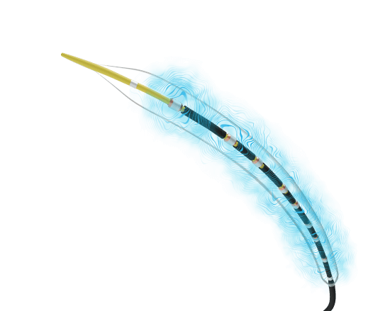
Shockwave IVL Above the Knee
Elevate outcomes: Shockwave Intravascular Lithotripsy (IVL) helps you maximize luminal gain, minimize complications and improve outcomes in calcified peripheral artery disease (PAD) above the knee.
Improving Outcomes Above the Knee (ATK) with Shockwave IVL
Shockwave IVL is an endovascular option that enables superior vessel preparation and provides excellent long-term results in the upper leg, overcoming historic challenges in treatment. For instance:
- Iliac occlusive disease treatment can lead to bleeding due to rupture and acute limb ischemia due to significant dissection1
- A significant number of patients with calcified CFA disease are not surgical candidates2, 3
- The popliteal artery is often considered a “no stent zone” due to being at a high flexion point4
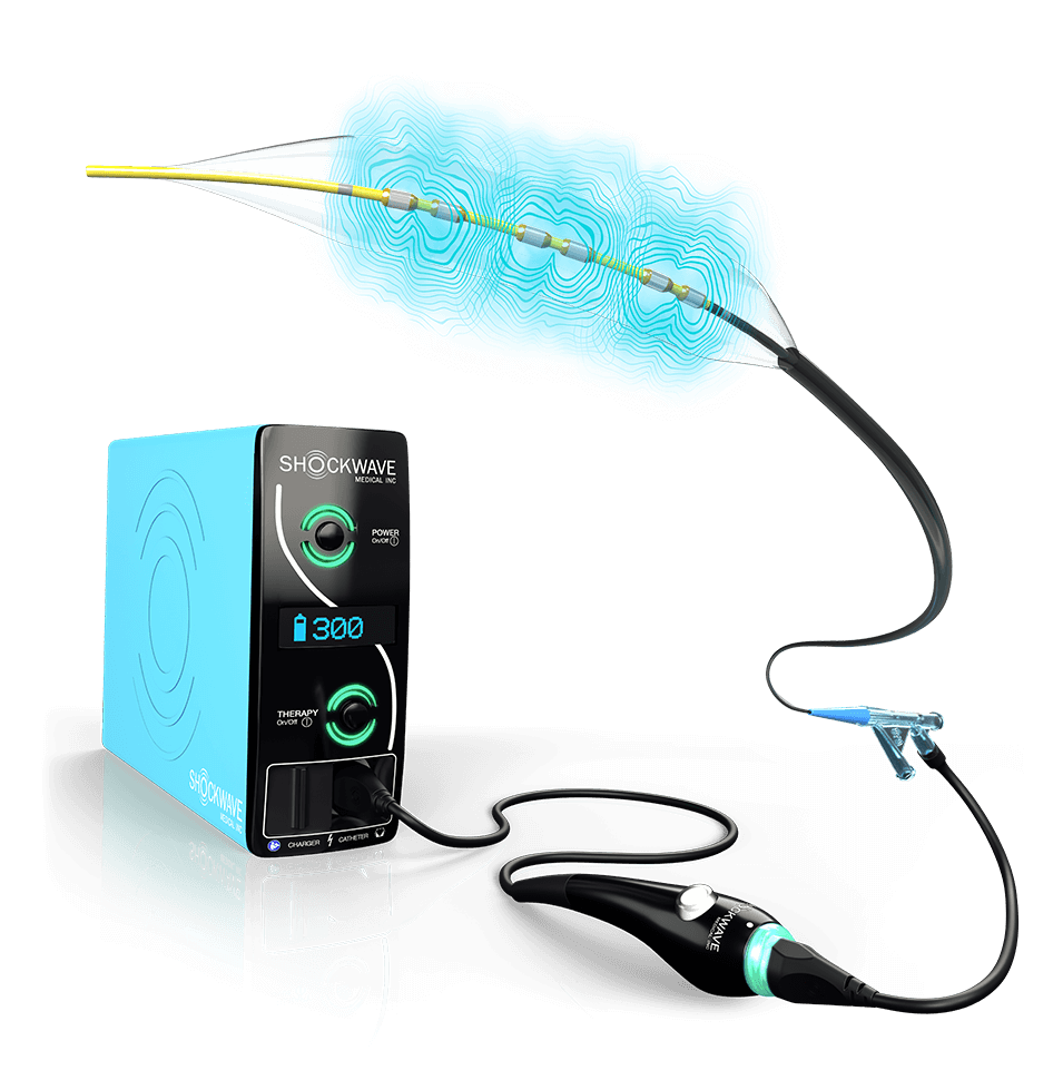
However, with Shockwave IVL’s purpose-built catheters, you can safely modify calcium at ultra-low pressure, thanks to a mechanism of action (MOA) that manages concerns of treating calcium in high-risk vessels.
1 Armstrong et. al. Cardiovascular Revascularization Medicine, 2020
2 Wong et al. CCI, 2018
3 GouëfficY et al,. JACC Cardiovasc Interv, 2017
4 Desyatova, Anastasia et al., Journal of the Royal Society Interface, 2017; 14(128)
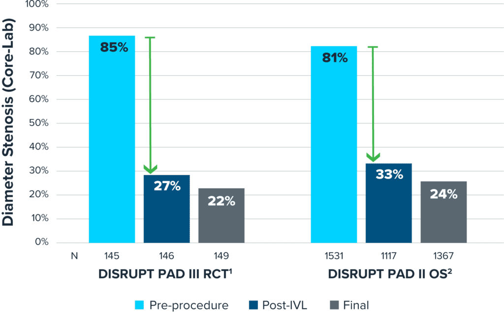
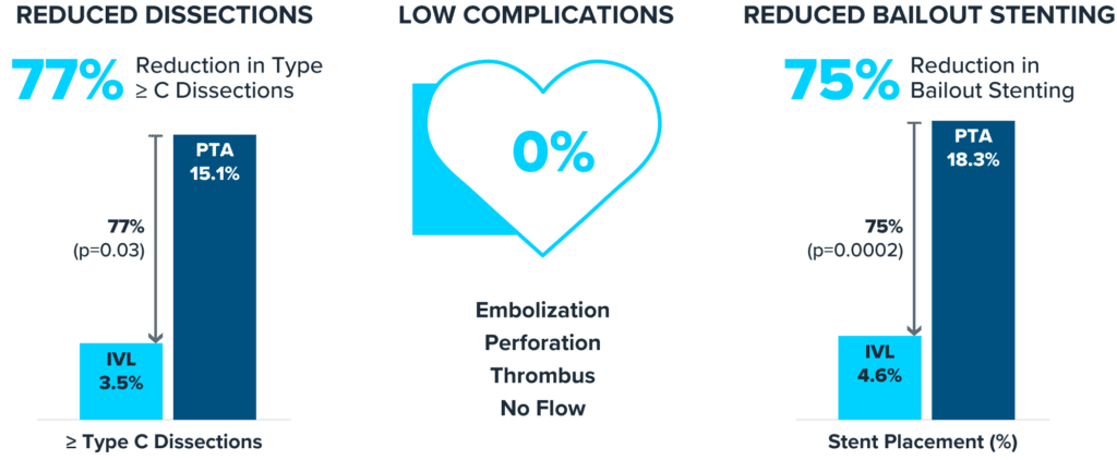
Shockwave IVL: Consistent Safety and Effectiveness
The DISRUPT PAD III Randomized Clinical Trial (RCT) and DISRUPT PAD III Observational Study (OS) showed exceptional safety profile and proven effective calcium modification.
Accordion Section
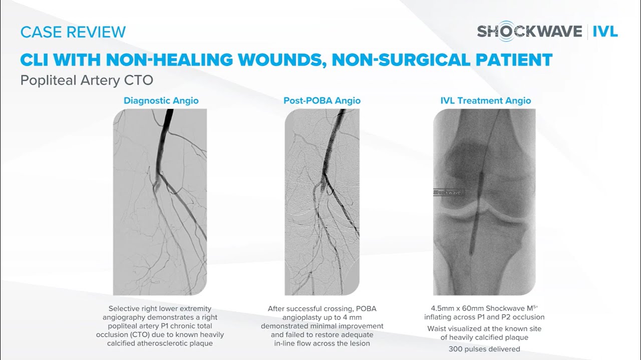
One Patient Story Shows the Possibilities
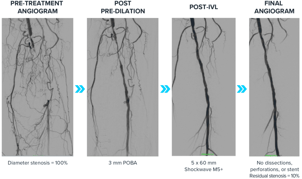
Treating a Calcified Popliteal Chronic Total Occlusion (CTO) with No Complications | Case courtesy of Dr. Anand Prasad
A 72-year-old female patient presented with lifestyle-limiting claudication and rest pain at night with worse symptoms in the left leg. The patient is a former smoker, has diabetes, CTO and chronic kidney disease (CKD). Invasive angiography of the left leg showed a chronic total occlusion in the P1 and P2 segments of the popliteal with no iliac disease and three vessel runoff to the feet. The CTO was crossed and a 3.0 mm POBA was used for the initial dilation. Then a 5.0 mm Shockwave M5+ was used along the calcified segments, finishing with a drug-coated balloon (DCB). Intravascular ultrasound (IVUS) imaging of calcified segments showed no dissections and good vessel expansion.
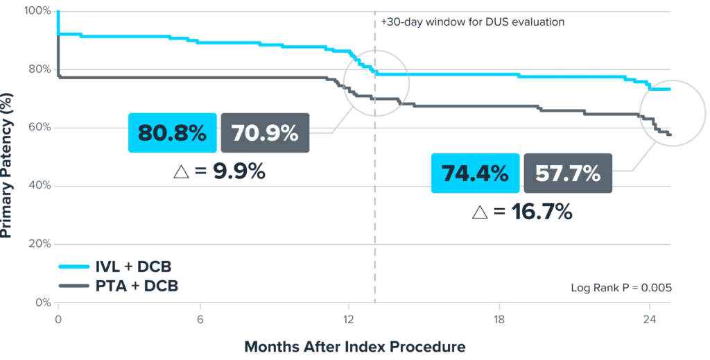
Shockwave IVL Offers Preserved Options and Excellent Long-Term Results1
Accordion Section

Featured Peripheral Case Reviews
View All Peripheral Case Reviews-

Dr. Raj Pyne Case Review: Popliteal CTO in Patient with CLTI
-
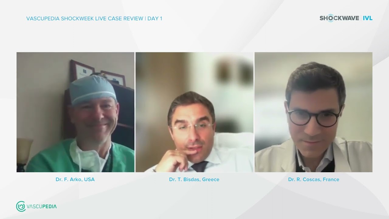
Shockwave IVL & Vascupedia Live Case Review Day 1: Tackling Calcified Disease in Large Vessels
-
Shockwave IVL in the Iliacs Discussion & Case Review
-
Intravascular Lithotripsy for Peripheral Artery Calcification: Mid-term Outcomes From the Randomized Disrupt PAD III Trial, Tepe et al, July 2022
-
Endovascular Intravascular Lithotripsy in the Treatment of Calcific Common Femoral Artery Disease: A Case Series With an 18-Month Follow-Up, Baig et al, October 2022