Dr. Raj Pyne is a paid consultant for Shockwave Medical.

Peripheral IVL  

Shockwave M5+, Shockwave M5, Shockwave S4, Shockwave L6 and Shockwave E8 Safety Information  

In the United States: Rx only.  

Indications for Use—The Shockwave Medical Intravascular Lithotripsy (IVL) System is intended for lithotripsy-enhanced balloon dilatation of lesions, including calcified lesions, in the peripheral vasculature, including the iliac, femoral, ilio-femoral, popliteal, infra-popliteal, and renal arteries. Not for use in the coronary, carotid or cerebral vasculature.  

Contraindications—Do not use if unable to pass 0.014″ (M5, M5+, S4, E8) or 0.018″ (L6) guidewire across the lesion-Not intended for treatment of in-stent restenosis or in coronary, carotid, or cerebrovascular arteries.

Warnings—Only to be used by physicians who are familiar with interventional vascular procedures—Physicians must be trained prior to use of the device—Use the generator in accordance with recommended settings as stated in the Operator’s Manual. 

Precautions—use only the recommended balloon inflation medium—Appropriate anticoagulant therapy should be administered by the physician—Decision regarding use of distal protection should be made based on physician assessment of treatment lesion morphology. 

Adverse effects–Possible adverse effects consistent with standard angioplasty include–Access site complications–Allergy to contrast or blood thinner–Arterial bypass surgery—Bleeding complications—Death—Fracture of guidewire or device—Hypertension/Hypotension—Infection/sepsis—Placement of a stent—renal failure—Shock/pulmonary edema—target vessel stenosis or occlusion—Vascular complications. Risks unique to the device and its use—Allergy to catheter material(s)— Device malfunction or failure—Excess heat at target site. 

Prior to use, please reference the Instructions for Use for more information on indications, contraindications, warnings, precautions and adverse events. www.shockwavemedical.com/ifu  

Dr. Peter O’Kane reviews a bifurcation left main case with eccentric calcification.


Dr. O’Kane is a paid consultant for Shockwave Medical.
Coronary IVL

Shockwave C2 and Shockwave C2+ Safety Information

In the United States: Rx only.

Indications for Use – The Shockwave Intravascular Lithotripsy (IVL) System with the Shockwave C2 and C2+ Coronary IVL Catheter is indicated for lithotripsy-enabled, low-pressure balloon dilatation of severely calcified, stenotic de novo coronary arteries prior to stenting.

Contraindications – The Shockwave C2 and C2+ Coronary IVL System is contraindicated for the following: This device is not intended for stent delivery. This device is not intended for use in carotid or cerebrovascular arteries.

Warnings – Use the IVL Generator in accordance with recommended settings as stated in the Operator’s Manual. The risk of a dissection or perforation is increased in severely calcified lesions undergoing percutaneous treatment, including IVL. Appropriate provisional interventions should be readily available. Balloon loss of pressure was associated with a numerical increase in dissection which was not statistically significant and was not associated with MACE. Analysis indicates calcium length is a predictor of dissection and balloon loss of pressure. IVL generates mechanical pulses which may cause atrial or ventricular capture in bradycardic patients. In patients with implantable pacemakers and defibrillators, the asynchronous capture may interact with the sensing capabilities. Monitoring of the electrocardiographic rhythm and continuous arterial pressure during IVL treatment is required. In the event of clinically significant hemodynamic effects, temporarily cease delivery of IVL therapy.

Precautions – Only to be used by physicians trained in angiography and intravascular coronary procedures. Use only the recommended balloon inflation medium. Hydrophilic coating to be wet only with normal saline or water and care must be taken with sharp objects to avoid damage to the hydrophilic coating. Appropriate anticoagulant therapy should be administered by the physician. Precaution should be taken when treating patients with previous stenting within 5mm of target lesion.

Potential adverse effects consistent with standard based cardiac interventions include- Abrupt vessel closure – Allergic reaction to contrast medium, anticoagulant and/or antithrombotic therapy-Aneurysm-Arrhythmia-Arteriovenous fistula-Bleeding complications-Cardiac tamponade or pericardial effusion-Cardiopulmonary arrest-Cerebrovascular accident (CVA)-Coronary artery/vessel occlusion, perforation, rupture or dissection-Coronary artery spasm-Death-Emboli (air, tissue, thrombus or atherosclerotic emboli)-Emergency or nonemergency coronary artery bypass surgery-Emergency or nonemergency percutaneous coronary intervention-Entry site complications-Fracture of the guide wire or failure/malfunction of any component of the device that may or may not lead to device embolism, dissection, serious injury or surgical intervention-Hematoma at the vascular access site(s)- Hemorrhage-Hypertension/Hypotension-Infection/sepsis/fever-Myocardial Infarction-Myocardial Ischemia or unstable angina-Pain-Peripheral Ischemia-Pseudoaneurysm-Renal failure/insufficiency-Restenosis of the treated coronary artery leading to revascularization-Shock/pulmonary edema-Slow flow, no reflow, or abrupt closure of coronary artery-Stroke Thrombus-Vessel closure, abrupt-Vessel injury requiring surgical repair-Vessel dissection, perforation, rupture, or spasm.

Risks identified as related to the device and its use: Allergic/immunologic reaction to the catheter material(s) or coating-Device malfunction, failure, or balloon loss of pressure leading to device embolism, dissection, serious injury or surgical intervention-Atrial or ventricular extrasystole-Atrial or ventricular capture.

Prior to use, please reference the Instructions for Use for more information on indications, contraindications, warnings, precautions and adverse events. www.shockwavemedical.com/IFU.

Treating Challenging Calcium Morphologies with the new Shockwave C2+

Hot off the press! Check out the latest Cardiovascular News supplement sponsored by Shockwave Medical.

This set of articles has a fresh perspective on today’s management of coronary calcium. It includes a deep dive into the use of intravascular lithotripsy for treating calcified nodules, and early experience with the new Shockwave C2+ coronary catheter.

Dr. Tom Johnson (University Hospitals Bristol & Weston NHS Foundation Trust, Bristol, UK) outlines how Shockwave IVL disrupted the landscape of UK interventional cardiology early in 2018. When introduced, it generated renewed enthusiasm for the management of complex calcific coronary disease and it impacted coronary calcium treatment in his centre over these last five years. Simplicity of use has been pivotal to make IVL the ‘go-to’ adjunctive tool for calcium modification.

The second article focuses on management of eccentric and nodular calcium, frequently present in CAC, associated with poor PCI outcomes and adverse long-term results. Dr. Nicolas Amabile (Institut Mutualiste Montsouris, Paris, France) discusses the best strategies to adequately manage these calcium morphologies and reports a case in which IVL was successfully used to treat calcified nodules in a challenging location.

Dr. Kambis Mashayekhi and his team at Herzzentrum Lahr/Baden (Lahr, Germany) detail their intravascular lithotripsy journey in the last article. They explain how the new Shockwave C2+ catheter can change the treatment of calcified coronary lesions and share a case study involving a diffuse calcified left main bifurcation.

Read the Publication


Drs. Spratt and Wilson are paid consultants for Shockwave Medical.

Coronary Important Safety Information:

In the United States: Rx only.

Indications for Use—The Shockwave Intravascular Lithotripsy (IVL) System with the Shockwave C2 Coronary IVL Catheter is indicated for lithotripsy-enabled, low-pressure balloon dilatation of severely calcified, stenotic de novo coronary arteries prior to stenting.

Contraindications—The Shockwave C2 Coronary IVL System is contraindicated for the following: This device is not intended for stent delivery. This device is not intended for use in carotid or cerebrovascular arteries.

Warnings— Use the IVL Generator in accordance with recommended settings as stated in the Operator’s Manual. The risk of a dissection or perforation is increased in severely calcified lesions undergoing percutaneous treatment, including IVL. Appropriate provisional interventions should be readily available. Balloon loss of pressure was associated with a numerical increase in dissection which was not statistically significant and was not associated with MACE.  Analysis indicates calcium length is a predictor of dissection and balloon loss of pressure.  IVL generates mechanical pulses which may cause atrial or ventricular capture in bradycardic patients. In patients with implantable pacemakers and defibrillators, the asynchronous capture may interact with the sensing capabilities. Monitoring of the electrocardiographic rhythm and continuous arterial pressure during IVL treatment is required.  In the event of clinically significant hemodynamic effects, temporarily cease delivery of IVL therapy.

Precautions— Only to be used by physicians trained in angiography and intravascular coronary procedures. Use only the recommended balloon inflation medium. Hydrophilic coating to be wet only with normal saline or water and care must be taken with sharp objects to avoid damage to the hydrophilic coating. Appropriate anticoagulant therapy should be administered by the physician. Precaution should be taken when treating patients with previous stenting within 5mm of target lesion.

Potential adverse effects consistent with standard based cardiac interventions include– Abrupt vessel closure – Allergic reaction to contrast medium, anticoagulant and/or antithrombotic therapy-Aneurysm-Arrhythmia-Arteriovenous fistula-Bleeding complications-Cardiac tamponade or pericardial effusion-Cardiopulmonary arrest-Cerebrovascular accident (CVA)-Coronary artery/vessel occlusion, perforation, rupture or dissection-Coronary artery spasm-Death-Emboli (air, tissue, thrombus or atherosclerotic emboli)-Emergency or non-emergency coronary artery bypass surgery-Emergency or non-emergency percutaneous coronary intervention-Entry site complications-Fracture of the guide wire or failure/malfunction of any component of the device that may or may not lead to device embolism, dissection, serious injury or surgical intervention-Hematoma at the vascular access site(s)-Hemorrhage-Hypertension/Hypotension-Infection/sepsis/fever-Myocardial Infarction-Myocardial Ischemia or unstable angina-Pain-Peripheral Ischemia-Pseudoaneurysm-Renal failure/insufficiency-Restenosis of the treated coronary artery leading to revascularization-Shock/pulmonary edema-Slow flow, no reflow, or abrupt closure of coronary artery-Stroke-Thrombus-Vessel closure, abrupt-Vessel injury requiring surgical repair-Vessel dissection, perforation, rupture, or spasm. Risks identified as related to the device and its use: Allergic/immunologic reaction to the catheter material(s) or coating-Device malfunction, failure, or balloon loss of pressure leading to device embolism, dissection, serious injury or surgical intervention-Atrial or ventricular extrasystole-Atrial or ventricular capture.

Prior to use, please reference the Instructions for Use for more information on warnings, precautions and adverse events.  https://shockwavemedical.com/IFU

Please contact your local Shockwave representative for specific country availability and refer to the Shockwave C2 instructions for use containing important safety information.

We sat down with Dr. Neel Butala, Interventional Cardiologist, University of Colorado Anschutz School of Medicine, University of Colorado Hospital after presenting the first results from his SCAI Early Career Research Award, “Use of Calcium Modification for Coronary Lesions after Introduction of Coronary IVL: National Trends and Hospital Variation” at SCAI2023 to get his perspective on the first data cut.

View Dr. Butala’s presentation here.


How has Shockwave coronary IVL impacted the calcium modification landscape?

Dr. Butala: Coronary IVL has dramatically changed how people treat coronary calcium. In a little over two years since initial approval in the United States, coronary IVL became the most frequently used calcium modification strategy for treatment of coronary lesions. Although it displaced other forms of calcium modification slightly, the spread of coronary IVL has generally led to an overall increase in use of calcium modification strategies. However, it is important to keep in mind that the dissemination of this technology has not been even. There is wide variation in use of calcium modification strategies as well as in use of coronary IVL in particular. As the landscape of calcium modification continues to evolve and this technology is introduced to more centers, I suspect that use of coronary IVL will continue to grow.


How would you interpret these findings – why is this so?

Dr. Butala: Our findings on trends in use of calcium modification strategies suggest that coronary IVL is truly a game changer. The speed with which coronary IVL has become the dominant calcium modification strategy speaks to the benefits of the technology and ease of use.


What are your key takeaways from this research project so far?

Dr. Butala: My key takeaways are that the introduction of coronary IVL has rapidly changed how physicians treat calcified coronary lesions and has led to greater use of calcium modification strategies overall. However, there is still wide variation in use of calcium modification strategies as well as coronary IVL and these are used in only a minority of cases at most centers.


How do these findings align with your current practice?

Dr. Butala: The findings make sense to me and reflect my practice. Since introduction of coronary IVL, my approach to calcified lesions has changed considerably. I am using IVL in many cases that would have previously required atherectomy, and I am also more comfortable taking on challenging lesions now that I have IVL in my toolbelt.


Were any of the findings surprising to you?

Dr. Butala: I was most surprised by how quickly IVL took over to become the most commonly used calcium strategy.


How do you anticipate the IC community to react to this data?

Dr. Butala: I think this will be in line with what many expected. I also think this will encourage more physicians to try coronary IVL if they haven’t already.


What should we look for next as you further dig into this topic?

Dr. Butala: The next step is for me to look at predictors of calcium modification strategy and IVL use to see if there are any disparities in care. Coronary IVL is a great case study in dissemination of a novel technology in the IC community, and it is important to investigate whether anyone was left behind. I also hope to update the trends and variation analyses with additional quarters of data and present it at a future meeting.


Dr. Neel Butala is a paid consultant for Shockwave Medical.

Coronary Important Safety Information:

In the United States: Rx only.

Indications for Use—The Shockwave Intravascular Lithotripsy (IVL) System with the Shockwave C2 Coronary IVL Catheter is indicated for lithotripsy-enabled, low-pressure balloon dilatation of severely calcified, stenotic de novo coronary arteries prior to stenting.

Contraindications—The Shockwave C2 Coronary IVL System is contraindicated for the following: This device is not intended for stent delivery. This device is not intended for use in carotid or cerebrovascular arteries.

Warnings— Use the IVL Generator in accordance with recommended settings as stated in the Operator’s Manual. The risk of a dissection or perforation is increased in severely calcified lesions undergoing percutaneous treatment, including IVL. Appropriate provisional interventions should be readily available. Balloon loss of pressure was associated with a numerical increase in dissection which was not statistically significant and was not associated with MACE.  Analysis indicates calcium length is a predictor of dissection and balloon loss of pressure.  IVL generates mechanical pulses which may cause atrial or ventricular capture in bradycardic patients. In patients with implantable pacemakers and defibrillators, the asynchronous capture may interact with the sensing capabilities. Monitoring of the electrocardiographic rhythm and continuous arterial pressure during IVL treatment is required.  In the event of clinically significant hemodynamic effects, temporarily cease delivery of IVL therapy.

Precautions— Only to be used by physicians trained in angiography and intravascular coronary procedures. Use only the recommended balloon inflation medium. Hydrophilic coating to be wet only with normal saline or water and care must be taken with sharp objects to avoid damage to the hydrophilic coating. Appropriate anticoagulant therapy should be administered by the physician. Precaution should be taken when treating patients with previous stenting within 5mm of target lesion.

Potential adverse effects consistent with standard based cardiac interventions include– Abrupt vessel closure – Allergic reaction to contrast medium, anticoagulant and/or antithrombotic therapy-Aneurysm-Arrhythmia-Arteriovenous fistula-Bleeding complications-Cardiac tamponade or pericardial effusion-Cardiopulmonary arrest-Cerebrovascular accident (CVA)-Coronary artery/vessel occlusion, perforation, rupture or dissection-Coronary artery spasm-Death-Emboli (air, tissue, thrombus or atherosclerotic emboli)-Emergency or non-emergency coronary artery bypass surgery-Emergency or non-emergency percutaneous coronary intervention-Entry site complications-Fracture of the guide wire or failure/malfunction of any component of the device that may or may not lead to device embolism, dissection, serious injury or surgical intervention-Hematoma at the vascular access site(s)-Hemorrhage-Hypertension/Hypotension-Infection/sepsis/fever-Myocardial Infarction-Myocardial Ischemia or unstable angina-Pain-Peripheral Ischemia-Pseudoaneurysm-Renal failure/insufficiency-Restenosis of the treated coronary artery leading to revascularization-Shock/pulmonary edema-Slow flow, no reflow, or abrupt closure of coronary artery-Stroke-Thrombus-Vessel closure, abrupt-Vessel injury requiring surgical repair-Vessel dissection, perforation, rupture, or spasm. Risks identified as related to the device and its use: Allergic/immunologic reaction to the catheter material(s) or coating-Device malfunction, failure, or balloon loss of pressure leading to device embolism, dissection, serious injury or surgical intervention-Atrial or ventricular extrasystole-Atrial or ventricular capture.

Prior to use, please reference the Instructions for Use for more information on warnings, precautions and adverse events.  https://shockwavemedical.com/IFU

Please contact your local Shockwave representative for specific country availability and refer to the Shockwave C2 instructions for use containing important safety information.

We asked Prof. James Spratt and Dr. Simon Wilson to share their IVL journey since the launch of Shockwave C2 five years ago and their initial experience with the new Shockwave C2+. Regardless of the physicians experience, the overall opinion is the same: “IVL is quick to learn, easy to use, very safe, and effective across a broad range of calcium types that other technologies can’t compete with. It has changed the landscape for calcium modification and the new Shockwave C2+ catheter continues to expand physicians ability to deal effectively and safely with severe calcium in a broader range of patients.” Read more below:

Shockwave Medical launched its coronary catheter, Shockwave C2, five years ago and you were one of the first consultants using IVL. Can you take us back to the beginning to the first time you used it and what your thoughts were?

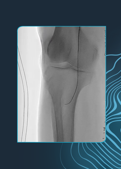
Prof. Spratt: With the initial launch, we were very curious to understand whether the product worked! Often where there is hype, it is swiftly followed by disappointment, but not in this case. Instrumental to our understanding of how and where IVL worked was our use of intravascular imaging. It was highly instructive to see fractures in previously rigid calcium and highly rewarding to get excellent results in challenging patients. Yet, despite the impressive efficacy, the safety profile is what reassured us to continue to explore what IVL could offer.

Your generation was once referred to as the “IVL generation.” Can you tell us about your experience with coronary intravascular lithotripsy (IVL) and how it influenced and shaped your treatment algorithm for calcified lesions?

Dr. Wilson: I was a keen user of atherectomy but quite often felt a bit disappointed with the outcome and never felt that cutting or high-pressure balloons were remotely suited to dealing with severe calcium. IVL has been hugely impactful to my practice. It is so easy to use, extremely safe, and just makes sense in terms of the mechanism of action. There is no doubt that having IVL available has allowed me to treat a much wider range of calcium types in a broader range of patients with a high degree of confidence in the devices safety and the final result.

How did IVL change your approach to the treatment of calcified lesions? Can you comment on your contemporary calcium algorithm compared to five years ago when IVL was introduced?

Prof. Spratt: Prior to the introduction of IVL, the options for treating patients with severe calcified coronary artery disease (CCAD) were limited. We frequently used rotational atherectomy and indeed continue to do so, but were aware of its limitations in failing to address deep calcium or indeed performing significant debulking. Following rota, all the other options were balloon-based, with the attendant limitations in nodular or eccentric calcium. This led to safety concerns over high-pressure balloon inflation & the risks of vessel perforation and ultimately led to less good clinical outcomes for patients. It is fair to say that IVL changed this landscape fundamentally & is now the treatment of choice for virtually all types of CCAD. Rotational atherectomy remains the treatment of choice for critical or uncrossable lesions, but beyond this, if the calcium burden is significant, IVL is the therapy of choice.

How would you describe the mechanism of action of IVL to your colleagues that are just starting with IVL? How does it compare with other specialty balloons?

Dr. Wilson: Non-compliant, cutting, and OPN balloons all work through applying hydrostatic force and are therefore not selective for calcium. On inflation, they will invariably expand away from resistance and with high-pressure inflations, risk major barotrauma. IVL as we know uses shockwaves that travel through the vessel wall generating disruptive forces only when calcium is encountered. IVL is therefore selective for calcium and can modify both superficial and deeper calcium as well as eccentric, concentric and nodular sub-types. Where there is significant calcium that has not resolved with an NC balloon at standard pressures, I no longer see an obvious role for anything other than IVL or atherectomy.

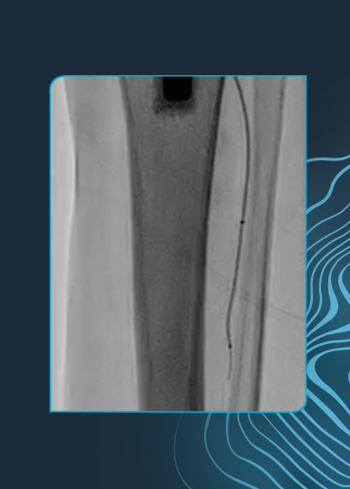
As one of the leading IVL centers in Europe, you had the chance to test first-hand the new Shockwave C2+ catheter with 50% more pulses. In general, what are your impressions of Shockwave C2+?

Prof. Spratt: We have been pleased. It extends the possibilities of IVL, enabling us to target more challenging lesions, such as nodular calcium, or longer lesions. There appears to be no durability or safety trade-off, so the access to 50% more “shocks” for the same cost is a win-win.

What case types or patient types that you believe the new Shockwave C2+ can better address compared to Shockwave C2?

Dr. Wilson: Broadly speaking, the impact of balloons relates to the hydrostatic force applied. Atherectomy depends on the contact area, whereas IVL can be thought of in terms of energy (or pulses) applied. The 50% increase in pulses gives you the confidence to deal with longer segments of severe calcium or two vessel calcific disease, where previously you might be reaching for atherectomy. I also find the extra pulses can be especially useful in thick eccentric calcium, as we know this type of calcium can often need a bit more energy to resolve.

Do you have any tips to share with people who are just starting to use IVL, particularly for the new catheter with 120 pulses?

Prof. Spratt: The first “tip” is to watch the Shockwave Calcium Masterclass series! This will provide a solid understanding of both the technology and lesion understanding. As with any technology, understanding the goals of therapy, in this case restoration of vessel compliance sufficient to allow full stent expansion, is critical. To properly integrate IVL into your practice, intravascular imaging is essential. The Masterclass Series, produced in partnership with Optima Education is foundational in this respect and highly recommended.

Can you explain what “pulse management” with IVL means and how do you think about applying pulses when you’re treating different types of calcium morphologies? Will your pulse management strategies change with 120 pulses?

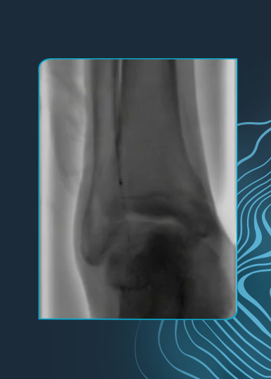
Dr. Wilson: To get the most out of IVL, it’s important to focus therapy on the most resistant segments. If it is a short segment of calcific disease, then it’s easy – put all the energy into that one spot. However, it’s very common to be dealing with much longer segments of calcific disease and although the C2+ gives you more margin, it’s still important to concentrate the majority of pulses into the most resistant segment or segments. I therefore make sure to understand where the most severe calcium on imaging is located angiographically and I almost always attempt pre-dilatation with a standard balloon ahead of IVL to understand where the greatest resistance is. Armed with this knowledge I then work from distal to proximal, covering the entire calcific segment with IVL but focusing the majority of pulses into the most resistant segments.

Will your calcium algorithm change with the introduction of Shockwave C2+? If so, can you take us through the changes?

Prof. Spratt: The most obvious iteration will be the ability to address longer sections of disease, or by targeting nodular or eccentric calcium. Important questions remain around what is the threshold of calcium that requires IVL; how best to identify an effective response, especially in nodular calcium and pulse management. We remain excited to explore these questions through research and education, what this space!


Drs. Spratt and Wilson are paid consultants for Shockwave Medical.

Coronary Important Safety Information:

In the United States: Rx only.

Indications for Use—The Shockwave Intravascular Lithotripsy (IVL) System with the Shockwave C2 Coronary IVL Catheter is indicated for lithotripsy-enabled, low-pressure balloon dilatation of severely calcified, stenotic de novo coronary arteries prior to stenting.

Contraindications—The Shockwave C2 Coronary IVL System is contraindicated for the following: This device is not intended for stent delivery. This device is not intended for use in carotid or cerebrovascular arteries.

Warnings— Use the IVL Generator in accordance with recommended settings as stated in the Operator’s Manual. The risk of a dissection or perforation is increased in severely calcified lesions undergoing percutaneous treatment, including IVL. Appropriate provisional interventions should be readily available. Balloon loss of pressure was associated with a numerical increase in dissection which was not statistically significant and was not associated with MACE.  Analysis indicates calcium length is a predictor of dissection and balloon loss of pressure.  IVL generates mechanical pulses which may cause atrial or ventricular capture in bradycardic patients. In patients with implantable pacemakers and defibrillators, the asynchronous capture may interact with the sensing capabilities. Monitoring of the electrocardiographic rhythm and continuous arterial pressure during IVL treatment is required.  In the event of clinically significant hemodynamic effects, temporarily cease delivery of IVL therapy.

Precautions— Only to be used by physicians trained in angiography and intravascular coronary procedures. Use only the recommended balloon inflation medium. Hydrophilic coating to be wet only with normal saline or water and care must be taken with sharp objects to avoid damage to the hydrophilic coating. Appropriate anticoagulant therapy should be administered by the physician. Precaution should be taken when treating patients with previous stenting within 5mm of target lesion.

Potential adverse effects consistent with standard based cardiac interventions include– Abrupt vessel closure – Allergic reaction to contrast medium, anticoagulant and/or antithrombotic therapy-Aneurysm-Arrhythmia-Arteriovenous fistula-Bleeding complications-Cardiac tamponade or pericardial effusion-Cardiopulmonary arrest-Cerebrovascular accident (CVA)-Coronary artery/vessel occlusion, perforation, rupture or dissection-Coronary artery spasm-Death-Emboli (air, tissue, thrombus or atherosclerotic emboli)-Emergency or non-emergency coronary artery bypass surgery-Emergency or non-emergency percutaneous coronary intervention-Entry site complications-Fracture of the guide wire or failure/malfunction of any component of the device that may or may not lead to device embolism, dissection, serious injury or surgical intervention-Hematoma at the vascular access site(s)-Hemorrhage-Hypertension/Hypotension-Infection/sepsis/fever-Myocardial Infarction-Myocardial Ischemia or unstable angina-Pain-Peripheral Ischemia-Pseudoaneurysm-Renal failure/insufficiency-Restenosis of the treated coronary artery leading to revascularization-Shock/pulmonary edema-Slow flow, no reflow, or abrupt closure of coronary artery-Stroke-Thrombus-Vessel closure, abrupt-Vessel injury requiring surgical repair-Vessel dissection, perforation, rupture, or spasm.

Risks identified as related to the device and its use: Allergic/immunologic reaction to the catheter material(s) or coating-Device malfunction, failure, or balloon loss of pressure leading to device embolism, dissection, serious injury or surgical intervention-Atrial or ventricular extrasystole-Atrial or ventricular capture.

Prior to use, please reference the Instructions for Use for more information on warnings, precautions and adverse events.  https://shockwavemedical.com/IFU

Please contact your local Shockwave representative for specific country availability and refer to the Shockwave C2 instructions for use containing important safety information.

Hear from Dr. JD Corl, interventional cardiologist at The Christ Hospital (Cincinnati, Ohio) on how Shockwave L6 is changing the game for large calcified peripheral vessels. 

In this video, Dr. Corl covers:  

  • The challenges and current endovascular options for treating large calcified peripheral vessels
  • A deep dive on the new Shockwave L6 catheter specifications and use-cases
  • A review of the first Shockwave L6 case (calcified bilateral iliacs)

All physicians are paid consultants of Shockwave Medical.
Important Safety Information
In the United States: Rx only.
Indications for Use—The Shockwave Medical Intravascular Lithotripsy (IVL) System is intended for lithotripsy-enhanced balloon dilatation of lesions, including calcified lesions, in the peripheral vasculature, including the iliac, femoral, ilio-femoral, popliteal, infra-popliteal, and renal arteries. Not for use in the coronary or cerebral vasculature.
Contraindications—Do not use if unable to pass 0.014″ (M5, M5+, S4) or 0.018″ (L6) guidewire across the lesion-Not intended for treatment of in-stent restenosis or in coronary, carotid, or cerebrovascular arteries.
Warnings—Only to be used by physicians who are familiar with interventional vascular procedures—Physicians must be trained prior to use of the device—Use the generator in accordance with recommended settings as stated in the Operator’s Manual.
Precautions—use only the recommended balloon inflation medium—Appropriate anticoagulant therapy should be administered by the physician—Decision regarding use of distal protection should be made based on physician assessment of treatment lesion morphology.
Adverse effects–Possible adverse effects consistent with standard angioplasty include–Access site complications–Allergy to contrast or blood thinner–Arterial bypass surgery—Bleeding complications—Death—Fracture of guidewire or device—Hypertension/Hypotension—Infection/sepsis—Placement of a stent—renal failure—Shock/pulmonary edema—target vessel stenosis or occlusion—Vascular complications.
Risks unique to the device and its use—Allergy to catheter material(s)— Device malfunction or failure—Excess heat at target site.
Prior to use, please reference the Instructions for Use for more information on indications, contraindications, warnings, precautions and adverse events. https://discover.shockwavemedical.com/ifu

Coronary IVL Benefits in Calcified Nodules Highlighted in JACC:CI

Coming off the heels of CRT23 where Dr. Richard Shlofmitz (St. Francis Heart Hospital, Long Island, NY) presented the longer-term data of coronary IVL in calcified nodules at two years, Dr. Ziad Ali (St. Francis Heart Hospital, Long Island, NY) et al. have recently published the initial coronary IVL experience in calcific nodules within JACC Cardiovascular Interventions. Following the publication, titled, “Safety and Effectiveness of Coronary Intravascular Lithotripsy for Treatment of Calcified Nodules,” we sat down with Dr. Ali, to learn more about this topical debate within the interventional community and separate the current perceived myths from the actual clinical evidence as it exists today in the treatment of calcified nodules.

Download JACC:CI Publication

Download CRT Presentation

Myth: Calcified nodules present most often in isolation

Clinical Evidence:

Although our eyes tend to focus on the “boulder” within the lumen of the coronary angiogram, in fact calcified nodules manifest in the presence of extensive severe calcification. Our data suggest that the burden of calcification is 4-fold higher in the presence of a nodule compared to without. In this regard it’s helpful to remember how calcified nodules actually form. As lipidic plaque accumulates within the artery wall, at a critical point the center of the lipid is so displaced from nutrient blood, that it develops into a necrotic core. To maintain vascular homeostasis, as a defense mechanism, the immune system can wall off the necrotic core by inducing calcification. This is one of the reasons that calcified nodules tend to occur in the proximal vessel – because the artery is large enough to accommodate a large lipid content through positive remodeling. This, now calcified former necrotic core, can either erupt into the vessel if the calcium is broken due to vessel torsion and bending, or simply protrude as the nodule becomes large enough or is pushed into the lumen by further lipid accumulation.


Myth: All calcified nodules are the same

Clinical Evidence:

Broadly speaking there are two types of calcified nodules. Eruptive calcified nodules, which are biologically active, and, protruding calcified nodules which are biologically inert. This is more than semantics, as the two types of nodules behave completely differently.

Eruptive calcified nodules “erupt” into the artery wall when a sheet of calcium breaks due to torsion, rotation and bending of the artery wall. Analogous to a glass in a bag, when the sheet calcium breaks some of the shards erupt into the vessel wall. These eruptions, which induce turbulent flow, and are devoid of endothelium, act as a nidus for thrombus formation causing up to 5% of all acute coronary syndromes.1

Protruding calcified nodules on the other hand, are as the name describe protrusions into the vessel wall. Analogous to a marbles in a bag, these protrusions either slowly grow into the lumen as they calcify or are pushed into the lumen by underlying lipid as lipid burden increases. While these protrusions can cause turbulent flow, they are covered by a fibrous cap and thus presumably endothelium, which does not induce thrombosis.

While these two different types of nodules are angiographically indistinguishable, intravascular imaging, in particular OCT, can help determine whether a nodule is eruptive or protruding. Recognizing this has direct clinical impact, as the response to therapy and the long term clinical outcomes of these two phenotypes is markedly different.


Myth: Atherectomy is the ideal tool for modifying nodular calcium

Clinical Evidence:

While it intuitively makes sense that shaving the nodule, or part thereof, would lead to the best acute result, data to support this theory is completely lacking. The only data to date comparing RA in calcified nodules versus non-nodular calcium showed a 3-fold higher clinically driven TLR at 5 years.2

As a parallel, treatment of calcified nodules with atherectomy could be compared to treatment of thrombus with aspiration catheters. While it intuitively makes sense, the volume of thrombus removed by aspiration has been shown to be less than 20%.3 Likewise, atherectomy only modifies a small proportion of the nodule, leaving the majority of the nodule, including the deep component unmodified. Moreover, because calcified nodules tend to occur in larger parts of the artery, the atherectomy may not impact the surrounding calcium due to wire-bias. On the contrary IVL, modifies the nodule by breaking it up into progressively smaller pieces with successive therapies. Moreover, since the IVL is wire-bias independent, it modifies not only the nodule itself, but the surrounding calcium, liberating circumferential vascular compliance.

The proof really is in the pudding. In our recent publication in JACC Intervention, we showed that the MSA, MLA and stent expansion were the same in severely calcified coronary arteries with or without nodular disease.4 That coupled with our recent CRT presentation, showing that the clinical outcomes comparing these two groups was similar, provides the strongest evidence base for interventional therapy of calcified nodules.


Myth: Nodular calcium is superficial; you just need to shave it off

Clinical Evidence:

Unfortunately, neither angiography, IVUS or OCT have the ability to determine the thickness of nodular calcium. Angiography lacks resolution, sound waves from IVUS reflect off the calcium, and OCT light is attenuated in the presence of very deep calcium or lipid. However, both conventional CT and micro-CT show that nodular calcification is deep. Pathophysiologically, by definition, nodular calcification has a deep component. As I mentioned earlier, nodules develop from necrotic core that progressively calcify, and necrotic cores only occur with positive remodeling vessel expansion and large lipid burdens.


Myth: Good, circumferential stent expansion with full apposition in calcified lesions equals a successful outcome

Clinical Evidence:

Again, while intuitively it makes sense that a circular stent would be optimal to restore vascular homeostasis, evidence to support this dogma is lacking. On the contrary neither eccentric stent expansion nor malapposition have been shown to impact clinical outcomes in DES. Blood really doesn’t care whether it goes through a circle or an oval, or behind a strut, or inside a strut. Rather, the most important determinant of long-term PCI outcome is the MSA. In keeping with this, in our recent publication in JACC Intervention, we showed that the MSA, MLA and stent expansion were the same in severely calcified coronary arteries with or without nodular disease. That coupled with our recent CRT presentation, showing that the clinical outcomes comparing these two groups was similar, provides the strongest evidence base for interventional therapy of calcified nodules.


Myth: A good acute outcome with calcified nodules is indicative of sustainable long-term outcomes

Clinical Evidence:

Sadly not. In a very recent analysis published in JACC Intervention we showed that the calcified nodule phenotype heavily impacts the long-term clinical outcome.5 Despite a better acute stent expansion in eruptive calcified nodules, this phenotype has a 4-fold higher risk of TLF compared to protruding nodules, reaching a 20% event rate at 2 years. It appears as though the mechanism by which the eruptive nodules fail, is a re-protrusion through the stent struts over time. Thus far we have not been able to determine which nodules are most likely to re-protrude other than those at hinge points in the vessel. Moreover, what treatment modality is optimal to prevent this re-protrusion remains unknown.


Myth: We know which treatment strategy is the ideal approach for calcific nodules today

Clinical Evidence:

What we do know is that the safety and effectiveness of IVL now with 2yr f/u in these patients is encouraging. But, there is still a lot we need to learn. Research in this area has been hampered by the prevalence of calcified nodules. Approximately 5-10% of severely calcified coronary lesions have a calcified nodule.6 Given this relatively low prevalence, it has proven challenging to perform prospective controlled studies. We need more real-world data in nodules across calcium arcs with intravascular imaging to start to determine the optimal algorithm for treatment of calcified nodules, such that we can improve both short and long term PCI outcomes. Certainly, the IVL data is encouraging and definitely proves a role for IVL in treatment of calcified nodules. Whether concomitant atherectomy may alter the plaque phenotype to facilitate more calcium fracture and lesion modification is certainly an interesting and valid hypothesis. We have embarked on a prospective study to evaluate this hypothesis, and hope to provide insight in the next few years.


1 Pengchata, P. et al. (2023). Characteristics and Pattern of Calcified Nodule and/or Nodular Calcification Detected by Intravascular Ultrasound on the Device-Oriented Composite Endpoint (DoCE) in Patients with Heavily Calcified Lesions Who Underwent Rotational Atherectomy-Assisted Percutaneous Coronary Intervention. Journal of Interventional Cardiology, 2023, 1–14. https://doi.org/10.1155/2023/6456695
2 Morofuji, T. et al. (2021). Clinical impact of calcified nodule in patients with heavily calcified lesions requiring rotational atherectomy. Catheterization and Cardiovascular Interventions, 97(1), 10–19. https://doi.org/10.1002/ccd.28896
3 Bhindi, R. et al. (2015). Culprit lesion thrombus burden after manual thrombectomy or percutaneous coronary intervention-alone in ST-segment elevation myocardial infarction: the optical coherence tomography sub-study of the TOTAL (ThrOmbecTomy versus PCI ALone) trial. European Heart Journal, 36(29), 1892–1900. https://doi.org/10.1093/eurheartj/ehv176
4 Ali, Z. A., et al. (2023). Safety and effectiveness of coronary intravascular lithotripsy for treatment of calcified nodules. JACC: Cardiovascular Interventions. https://doi.org/10.1016/j.jcin.2023.02.015
5 Sato T. et al. (2023). Impact of Eruptive vs Noneruptive Calcified Nodule Morphoplogy on Acuet and Long-Term Outcomes After Stenting. JACC: Cardiovascular Interventions. In Press.
6 Petrossian, G., et al. (2022). Role of Intracoronary Imaging in Acute Coronary Syndromes. US Cardiology Review, 16. https://doi.org/10.15420/usc.2022.03


Dr. Sandeep Basavarajaiah and Dr. Alfonso Ielasi are paid consultants of Shockwave Medical.


Coronary Important Safety Information:

In the United States: Rx only.

Indications for Use—The Shockwave Intravascular Lithotripsy (IVL) System with the Shockwave C2 Coronary IVL Catheter is indicated for lithotripsy-enabled, low-pressure balloon dilatation of severely calcified, stenotic de novo coronary arteries prior to stenting.

Contraindications—The Shockwave C2 Coronary IVL System is contraindicated for the following: This device is not intended for stent delivery. This device is not intended for use in carotid or cerebrovascular arteries.

Warnings— Use the IVL Generator in accordance with recommended settings as stated in the Operator’s Manual. The risk of a dissection or perforation is increased in severely calcified lesions undergoing percutaneous treatment, including IVL. Appropriate provisional interventions should be readily available. Balloon loss of pressure was associated with a numerical increase in dissection which was not statistically significant and was not associated with MACE.  Analysis indicates calcium length is a predictor of dissection and balloon loss of pressure.  IVL generates mechanical pulses which may cause atrial or ventricular capture in bradycardic patients. In patients with implantable pacemakers and defibrillators, the asynchronous capture may interact with the sensing capabilities. Monitoring of the electrocardiographic rhythm and continuous arterial pressure during IVL treatment is required.  In the event of clinically significant hemodynamic effects, temporarily cease delivery of IVL therapy.

Precautions— Only to be used by physicians trained in angiography and intravascular coronary procedures. Use only the recommended balloon inflation medium. Hydrophilic coating to be wet only with normal saline or water and care must be taken with sharp objects to avoid damage to the hydrophilic coating. Appropriate anticoagulant therapy should be administered by the physician. Precaution should be taken when treating patients with previous stenting within 5mm of target lesion.

Potential adverse effects consistent with standard based cardiac interventions include– Abrupt vessel closure – Allergic reaction to contrast medium, anticoagulant and/or antithrombotic therapy-Aneurysm-Arrhythmia-Arteriovenous fistula-Bleeding complications-Cardiac tamponade or pericardial effusion-Cardiopulmonary arrest-Cerebrovascular accident (CVA)-Coronary artery/vessel occlusion, perforation, rupture or dissection-Coronary artery spasm-Death-Emboli (air, tissue, thrombus or atherosclerotic emboli)-Emergency or non-emergency coronary artery bypass surgery-Emergency or non-emergency percutaneous coronary intervention-Entry site complications-Fracture of the guide wire or failure/malfunction of any component of the device that may or may not lead to device embolism, dissection, serious injury or surgical intervention-Hematoma at the vascular access site(s)-Hemorrhage-Hypertension/Hypotension-Infection/sepsis/fever-Myocardial Infarction-Myocardial Ischemia or unstable angina-Pain-Peripheral Ischemia-Pseudoaneurysm-Renal failure/insufficiency-Restenosis of the treated coronary artery leading to revascularization-Shock/pulmonary edema-Slow flow, no reflow, or abrupt closure of coronary artery-Stroke-Thrombus-Vessel closure, abrupt-Vessel injury requiring surgical repair-Vessel dissection, perforation, rupture, or spasm. Risks identified as related to the device and its use: Allergic/immunologic reaction to the catheter material(s) or coating-Device malfunction, failure, or balloon loss of pressure leading to device embolism, dissection, serious injury or surgical intervention-Atrial or ventricular extrasystole-Atrial or ventricular capture.

Prior to use, please reference the Instructions for Use for more information on warnings, precautions and adverse events.  https://shockwavemedical.com/IFU

Please contact your local Shockwave representative for specific country availability and refer to the Shockwave C2 instructions for use containing important safety information.

Learn How Coronary IVL is Currently Utilized Within Non-Surgical Facilities

Want to learn more about Coronary IVL’s place within existing facilities lacking surgical cardiac backup? Cardiovascular Business and Shockwave Medical is excited to present this on demand webinar titled: No Surgical Backup? No Problem. This webinar consists of an expert panel who provide real-world insights and implications of Coronary IVL usage within non-surgical facilities. Topics within this event include a review of the recent SCAI Expert Consensus Document regarding PCI within non-surgical facilities, an introduction to Shockwave IVL, and a real-world clinical and administrational perspective of Coronary IVL within an existing non-surgical facility. A fantastic roundtable discussion and Q+A close the webinar with additional insights regarding Coronary IVL within these important facilities.


Drs. Alex Truesdell, Poonam Velagapudi, Clay Sizemore, and Chris Newman are paid consultants for Shockwave Medical.

Important Safety Information

Please contact your local Shockwave representative for specific country availability and refer to the Shockwave C2 instructions for use containing important safety information.

Rx only

Indications for Use—The Shockwave Intravascular Lithotripsy (IVL) System with the Shockwave C2 Coronary IVL Catheter is indicated for lithotripsy-enabled, low-pressure balloon dilatation of severely calcified, stenotic de novo coronary arteries prior to stenting.

Contraindications—The Shockwave C2 Coronary IVL System is contraindicated for the following: This device is not intended for stent delivery. This device is not intended for use in carotid or cerebrovascular arteries.

Warnings— Use the IVL Generator in accordance with recommended settings as stated in the Operator’s Manual. The risk of a dissection or perforation is increased in severely calcified lesions undergoing percutaneous treatment, including IVL. Appropriate provisional interventions should be readily available. Balloon loss of pressure was associated with a numerical increase in dissection which was not statistically significant and was not associated with MACE.  Analysis indicates calcium length is a predictor of dissection and balloon loss of pressure.  IVL generates mechanical pulses which may cause atrial or ventricular capture in bradycardic patients. In patients with implantable pacemakers and defibrillators, the asynchronous capture may interact with the sensing capabilities. Monitoring of the electrocardiographic rhythm and continuous arterial pressure during IVL treatment is required.  In the event of clinically significant hemodynamic effects, temporarily cease delivery of IVL therapy.

Precautions— Only to be used by physicians trained in angiography and intravascular coronary procedures. Use only the recommended balloon inflation medium. Hydrophilic coating to be wet only with normal saline or water and care must be taken with sharp objects to avoid damage to the hydrophilic coating. Appropriate anticoagulant therapy should be administered by the physician. Precaution should be taken when treating patients with previous stenting within 5mm of target lesion.

Potential adverse effects consistent with standard based cardiac interventions include– Abrupt vessel closure – Allergic reaction to contrast medium, anticoagulant and/or antithrombotic therapy-Aneurysm-Arrhythmia-Arteriovenous fistula-Bleeding complications-Cardiac tamponade or pericardial effusion-Cardiopulmonary arrest-Cerebrovascular accident (CVA)-Coronary artery/vessel occlusion, perforation, rupture or dissection-Coronary artery spasm-Death-Emboli (air, tissue, thrombus or atherosclerotic emboli)-Emergency or non-emergency coronary artery bypass surgery-Emergency or non-emergency percutaneous coronary intervention-Entry site complications-Fracture of the guide wire or failure/malfunction of any component of the device that may or may not lead to device embolism, dissection, serious injury or surgical intervention-Hematoma at the vascular access site(s)-Hemorrhage-Hypertension/Hypotension-Infection/sepsis/fever-Myocardial Infarction-Myocardial Ischemia or unstable angina-Pain-Peripheral Ischemia-Pseudoaneurysm-Renal failure/insufficiency-Restenosis of the treated coronary artery leading to revascularization-Shock/pulmonary edema-Slow flow, no reflow, or abrupt closure of coronary artery-Stroke-Thrombus-Vessel closure, abrupt-Vessel injury requiring surgical repair-Vessel dissection, perforation, rupture, or spasm.

Risks identified as related to the device and its use: Allergic/immunologic reaction to the catheter material(s) or coating-Device malfunction, failure, or balloon loss of pressure leading to device embolism, dissection, serious injury or surgical intervention-Atrial or ventricular extrasystole-Atrial or ventricular capture.

Prior to use, please reference the Instructions for Use for more information on warnings, precautions and adverse events.  https://shockwavemedical.com/IFU

Prof. Michael Haude and Dr. Milad Golabkesh discuss the new Shockwave C2+ Coronary IVL catheter and share a complex PCI case.


Prof Haude is a paid consultant of Shockwave Medical.

Shockwave C2 and Shockwave C2+ Safety Information

In the United States: Rx only

Indications for Use— The Shockwave Intravascular Lithotripsy (IVL) System with the Shockwave C2 and C2+ Coronary IVL Catheter is indicated for lithotripsy-enabled, low-pressure balloon dilatation of severely calcified, stenotic de novo coronary arteries prior to stenting.

Contraindications— The Shockwave C2 and C2+ Coronary IVL System is contraindicated for the following: This device is not intended for stent delivery. This device is not intended for use in carotid or cerebrovascular arteries.

Warnings— Use the IVL Generator in accordance with recommended settings as stated in the Operator’s Manual. The risk of a dissection or perforation is increased in severely calcified lesions undergoing percutaneous treatment, including IVL. Appropriate provisional interventions should be readily available. Balloon loss of pressure was associated with a numerical increase in dissection which was not statistically significant and was not associated with MACE. Analysis indicates calcium length is a predictor of dissection and balloon loss of pressure. IVL generates mechanical pulses which may cause atrial or ventricular capture in bradycardic patients. In patients with implantable pacemakers and defibrillators, the asynchronous capture may interact with the sensing capabilities. Monitoring of the electrocardiographic rhythm and continuous arterial pressure during IVL treatment is required. In the event of clinically significant hemodynamic effects, temporarily cease delivery of IVL therapy.

Precautions— Only to be used by physicians trained in angiography and intravascular coronary procedures. Use only the recommended balloon inflation medium. Hydrophilic coating to be wet only with normal saline or water and care must be taken with sharp objects to avoid damage to the hydrophilic coating. Appropriate anticoagulant therapy should be administered by the physician. Precaution should be taken when treating patients with previous stenting within 5mm of target lesion.

Potential adverse effects consistent with standard based cardiac interventions include– Abrupt vessel closure – Allergic reaction to contrast medium, anticoagulant and/or antithrombotic therapy-Aneurysm-Arrhythmia-Arteriovenous fistula-Bleeding complications-Cardiac tamponade or pericardial effusion-Cardiopulmonary arrest-Cerebrovascular accident (CVA)-Coronary artery/vessel occlusion, perforation, rupture or dissection-Coronary artery spasm-Death-Emboli (air, tissue, thrombus or atherosclerotic emboli)-Emergency or nonemergency coronary artery bypass surgery-Emergency or nonemergency percutaneous coronary intervention-Entry site complications-Fracture of the guide wire or failure/malfunction of any component of the device that may or may not lead to device embolism, dissection, serious injury or surgical intervention-Hematoma at the vascular access site(s)- Hemorrhage-Hypertension/Hypotension-Infection/sepsis/fever-Myocardial Infarction-Myocardial Ischemia or unstable angina-Pain-Peripheral Ischemia-Pseudoaneurysm-Renal failure/insufficiency-Restenosis of the treated coronary artery leading to revascularization-Shock/pulmonary edema-Slow flow, no reflow, or abrupt closure of coronary artery-Stroke Thrombus-Vessel closure, abrupt-Vessel injury requiring surgical repair-Vessel dissection, perforation, rupture, or spasm.

Risks identified as related to the device and its use: Allergic/immunologic reaction to the catheter material(s) or coating-Device malfunction, failure, or balloon loss of pressure leading to device embolism, dissection, serious injury or surgical intervention-Atrial or ventricular extrasystole-Atrial or ventricular capture.

Prior to use, please reference the Instructions for Use for more information on indications, contraindications, warnings, precautions and adverse events. www.shockwavemedical.com/IFU.