Perspective on IVL-Induced Ventricular Capture from Keith D. Dawkins, M.D., Chief Medical Officer, Shockwave Medical, Inc.:

Late last week, one of our customers in the UK reported in the European Heart Journal what appears to be the first reported case of ventricular fibrillation (VF) associated with IVL. We appreciate that this case report was published in order for physicians to be informed of this rare event.

VF associated with IVL use is rare based on our clinical and commercial experience: this is the first report we are aware of in over 25K coronary IVL cases performed to date.

The report describes in detail how, in an off-label case, a severely calcified in-stent restenosis of the right coronary artery (RCA), IVL spikes fell on a T-wave, which then in-turn seems to have induced VF.

For background, IVL produces a low amount of mechanical energy, which is capable of inducing premature ventricular beats in primarily bradycardic patients, known as ‘IVL-induced ventricular capture,’ which was described well in the EuroIntervention publication from Wilson et al who documented its first report. It is important to understand that no electrical current leaves the IVL catheter. Rather, a small amount of mechanical energy is transferred to the vessel wall when sonic pressure waves are created that have been shown to create a stretch activated response in the myocardium.

Last month, the DISRUPT CAD III clinical trial investigators published in JACC the first prospective analysis on this topic as part of the 431-patient U.S. IDE study, which showed IVL-induced capture was common but benign, with the authors noting, “Decreased systolic blood pressure during the IVL procedure was more frequent in patients with IVL-induced capture compared to those without (40.5% vs. 24.5%; p = 0.0007). However, the magnitude of the drop in systolic blood pressure was similar between the 2 groups (18.9 vs. 23.5 mmHg systolic; p = 0.07). IVL-induced capture did not result in sustained ventricular arrhythmias during or immediately after the IVL procedure in any patient and was not associated with adverse events.”

While acknowledging that VF can occur in any case, diagnostic or interventional, a recently published report showed that the risk of VT or VF is 0.8% in diagnostic angiography, 1.1% for stable PCI and as much as 4.3% for primary PCI in AMI. In these cases, VF may also be caused by ischemic events, for example prolonged balloon inflations, not related to IVL.

VF is typically reversible with cardioversion in the cath lab, as was the case in this published report.

We understand IVL-induced ventricular capture, and while we have not heard of any other VF reports, we will continue to monitor this topic and encourage our customers to continue to report complaints to our quality team at [email protected].


Important Safety Information – Coronary IVL

Caution: In the United States, Shockwave C2 Coronary IVL catheters are investigational devices, limited by United States law to investigational use. DISRUPT CAD III Study

Shockwave C2 Coronary IVL catheters are commercially available in certain countries outside the U.S. Please contact your local Shockwave representative for specific country availability. The Shockwave C2 Coronary IVL catheters are indicated for lithotripsy-enhanced, low-pressure balloon dilatation of calcified, stenotic de novo coronary arteries prior to stenting. For the full IFU containing important safety information please visit: https://shockwavemedical.com/clinicians/international/coronary/shockwave-c2/

Following its presentation as a late-breaking clinical at TCT Connect and simultaneous publication in the Journal of the American College of Cardiology (JACC) we sat down with Dr. Jonathan Hill, Consultant Cardiologist at the Royal Brompton Hospital in London, to get his perspective on the results, how they build on DISRUPT CAD I and II outcomes, and where they fit into clinician’s daily decision-making process when treating calcified lesions. We hope you enjoy his perspective.


What does DISRUPT CAD III add to the existing results from DISRUPT CAD I and DISRUPT CAD II?

Dr. Hill: CAD III is the culmination of four years of research in over 600 patients that has spanned three studies. Its results are noteworthy in that it’s the first study that has been appropriately powered to evaluate the safety and effectiveness of IVL, which is important when evaluating low frequency angiographic complications, such as perforations and dissections, as well as clinical events, such as MI and TVR. It also is the first IVL study to prospectively look at IVL’s impact on heart rhythm, and follow patients for two years. Finally, we studied more patients with OCT than CAD I and II combined. For all of these reasons, its results have unveiled a significant amount of new insights about the therapy.


How would you summarize the key findings from DISRUPT CAD III?

Dr. Hill: There are four key findings from my perspective. First, CAD III successfully met both its primary safety and effectiveness endpoints, despite having one of the most challenging cohort of calcified lesions ever studied. Second, coronary IVL prior to DES implantation was well tolerated with a low rate of major peri-procedural clinical and angiographic complications, similar to what we found in CAD I and II. Third, IVL-induced ventricular capture was common, but was transient and benign with no clinical sequelae in any patient. And lastly, although this study represents the initial coronary IVL experience for U.S. operators, similar high procedural success and device crossing success, as well as low MACE and angiographic complications were achieved in both the initial roll-in cases and the patients included in the ITT analysis, demonstrating the relative ease of use of IVL for the first time in a study.


Why do you say that this was one of the most challenging cohorts of calcified lesions ever studied?

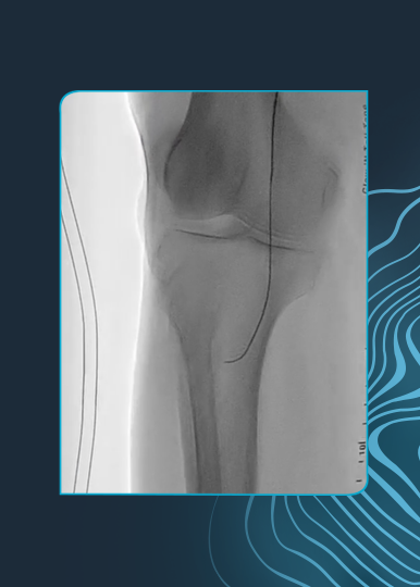
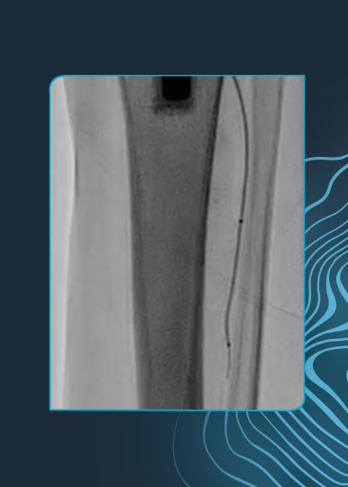
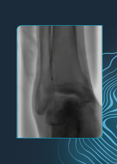
Dr. Hill: Following the ORBIT II design as our predicate study, the inclusion criteria for CAD III was designed to focus on finding the most severely calcified patients, only including patients with angiographic evidence of calcium on both sides of the artery, or more than 270 degrees of calcium under intravascular imaging. We were successful to that end, enrolling 100% of patients with severe calcium. The average calcium lesion length was very long at 47.9 mm, and the average calcium arc was 292.5 degrees with a thickness of 0.96 mm at the site of maximum calcification as measured by OCT. If you’re familiar with the rule of 5s with OCT, you’ll know that these are the most challenging type of calcified lesions to treat: thick, circumferential and diffuse calcium.


Were there any surprises or unexpected findings from DISRUPT CAD III?

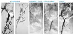
Dr. Hill: One of the current limitations of the technology has been its higher crossing profile when compared to a normal NC balloon. However, despite the marked severity of the calcified lesions treated, IVL was able to cross and deliver therapy in 98.2% of lesions, or 377 of 384 total patients, which then lead to a 99% rate of stent deliverability. I was surprised to see such a high crossing rate, especially considering that this was U.S. operators first use of the technology.

Another unexpected finding was related to fracture evidence under OCT. Interestingly, OCT showed excellent stent expansion in those lesions with and without calcium fractures identified by OCT despite the marked severity of the calcified lesions treated, meaning that IVL led to improved vessel compliance and favorable stent expansion even without demonstrable calcium fracture by OCT. I think that finding will surprise my fellow colleagues who use OCT regularly and have trained themselves to look for fractures to know if they have delivered enough energy to adequately prepare the artery for stent implantation. This raises the prospect of IVL induced microfractures not visible by OCT as an additional mechanism of increasing vessel compliance.


You mentioned IVL-induced ventricular capture was investigated prospectively in DISRUPT CAD III for the first time – what were the conclusions regarding this topic?

Dr. Hill: As many have seen in their own use with the technology, as well as what has been reported previously about IVL-induced ventricular capture in the EuroIntervention publication, CAD III found that this phenomenon was common, occurring in just over 40% of cases. Importantly, no serious adverse clinical events occurred as a result of IVL-induced capture. It was significantly more common if the pre-procedure heart rate was lower, particularly by multivariate analysis less than 60BPM. The drop in systolic blood pressure was more frequent, but not more severe, based on this analysis. And most important this was a transient phenomenon which resolved once IVL delivery stopped without significant sequalae.


What are your main takeaways from the OCT sub-study?

Dr. Hill: I think my main takeaways from the OCT sub-study would be that with 100 OCT patients studied, we have the largest analysis yet that provides us with new evidence that confirms and extends prior findings on IVL’s unique mechanism of action that enables optimal stent delivery, expansion and apposition. A few of the stats that I thought were interesting were the final MSA of 6.5 mm, which is a great outcome, the final stent expansion of 102% at the site of maximum calcification (where the calcium angle was 292° and 0.96 mm thick on average), and that post-IVL calcium fractures were observed in multiple longitudinal planes in 67% of lesions.

Looking specifically at the fractures, four correlative findings were that the minimum calcium angle that resulted in calcium fracture was 192° and the result already mentioned that regardless of calcium fracture identification by OCT, there were no differences between lesions with or without fractures in relation to the MSA, final area stenosis, and stent expansion. The fractures were also an average depth of 0.5 mm deep and 0.5 mm wide, which expanded to 1.3 mm after stent implantation.


How does DISRUPT CAD III change your use of IVL or your daily practice of calcium management?

Dr. Hill: I think there are several practice-changing finds for operators across Europe who have been using the technology for some time now. First, we know what to expect now with IVL-induced capture, including how often it occurs, in which scenarios and what impact it has on the patients so that we can mitigate it successfully. Two, when looking for post-IVL fractures for those who use OCT, the CAD III data would suggest that evidence of fracture is not a necessary indicator of adequate lesion preparation with IVL.

For those individuals who do not use intravascular imaging in everyday practice, this data shows that overall balloon expansion under angiography may be an alternative way to determine if the calcium has been modified adequately and that the vessel compliance has changed sufficiently for DES implantation.


Where do we go from here in regard to clinical evidence with IVL?

Dr. Hill: The study of IVL across CAD I, CAD II and CAD III provides a robust clinical data set of over 600 patients, and from the CAD III study, we have identified additional areas of interest beyond what was observed in the initial studies. First, I think it’s important to note that the JACC publication is the 30-day data, and these patients will be followed out to two years, which will be important as this will be the longest and most rigorous follow-up conducted on coronary IVL patients to date. Second, the fact that MSA, area stenosis, and stent expansion outcomes were excellent regardless of calcium fracture visualization by OCT is a novel finding and challenges the accepted practice of visual confirmation when using calcium modification tools. It may represent OCT’s limitation to detect subtle micro-fractures or out-of-plane fractures in calcified plaque, but this is just a hypothesis and we need to further assess. Third, while this patient population consisted of mostly lesions with circumferential calcium, further assessment of the OCT patients treated within the DISRUPT CAD program, which should be nearly 250 patients, will provide more insights regarding the similar performance in concentric & eccentric lesions that was previously found in the CAD II angiography-based study. And finally, addressing the major limitations of nearly all calcium modifications algorithms that are coming to light (with none based on data), randomized data across multiple calcium tools may be warranted. I have no doubt that RCTs will be done that include IVL and other calcium tools; in fact there are several smaller RCT – some investigator-sponsored and others independent – that have been initiated, and I look forward to their outcomes.


Dr. Jonathan Hill is a paid consultant of Shockwave Medical.

Coronary Important Safety Information:

In the United States: Rx only.

Indications for Use—The Shockwave Intravascular Lithotripsy (IVL) System with the Shockwave C2 Coronary IVL Catheter is indicated for lithotripsy-enabled, low-pressure balloon dilatation of severely calcified, stenotic de novo coronary arteries prior to stenting.

Contraindications—The Shockwave C2 Coronary IVL System is contraindicated for the following: This device is not intended for stent delivery. This device is not intended for use in carotid or cerebrovascular arteries.

Warnings— Use the IVL Generator in accordance with recommended settings as stated in the Operator’s Manual. The risk of a dissection or perforation is increased in severely calcified lesions undergoing percutaneous treatment, including IVL. Appropriate provisional interventions should be readily available. Balloon loss of pressure was associated with a numerical increase in dissection which was not statistically significant and was not associated with MACE.  Analysis indicates calcium length is a predictor of dissection and balloon loss of pressure.  IVL generates mechanical pulses which may cause atrial or ventricular capture in bradycardic patients. In patients with implantable pacemakers and defibrillators, the asynchronous capture may interact with the sensing capabilities. Monitoring of the electrocardiographic rhythm and continuous arterial pressure during IVL treatment is required.  In the event of clinically significant hemodynamic effects, temporarily cease delivery of IVL therapy.

Precautions— Only to be used by physicians trained in angiography and intravascular coronary procedures. Use only the recommended balloon inflation medium. Hydrophilic coating to be wet only with normal saline or water and care must be taken with sharp objects to avoid damage to the hydrophilic coating. Appropriate anticoagulant therapy should be administered by the physician. Precaution should be taken when treating patients with previous stenting within 5mm of target lesion.

Potential adverse effects consistent with standard based cardiac interventions include– Abrupt vessel closure – Allergic reaction to contrast medium, anticoagulant and/or antithrombotic therapy-Aneurysm-Arrhythmia-Arteriovenous fistula-Bleeding complications-Cardiac tamponade or pericardial effusion-Cardiopulmonary arrest-Cerebrovascular accident (CVA)-Coronary artery/vessel occlusion, perforation, rupture or dissection-Coronary artery spasm-Death-Emboli (air, tissue, thrombus or atherosclerotic emboli)-Emergency or non-emergency coronary artery bypass surgery-Emergency or non-emergency percutaneous coronary intervention-Entry site complications-Fracture of the guide wire or failure/malfunction of any component of the device that may or may not lead to device embolism, dissection, serious injury or surgical intervention-Hematoma at the vascular access site(s)-Hemorrhage-Hypertension/Hypotension-Infection/sepsis/fever-Myocardial Infarction-Myocardial Ischemia or unstable angina-Pain-Peripheral Ischemia-Pseudoaneurysm-Renal failure/insufficiency-Restenosis of the treated coronary artery leading to revascularization-Shock/pulmonary edema-Slow flow, no reflow, or abrupt closure of coronary artery-Stroke-Thrombus-Vessel closure, abrupt-Vessel injury requiring surgical repair-Vessel dissection, perforation, rupture, or spasm. Risks identified as related to the device and its use: Allergic/immunologic reaction to the catheter material(s) or coating-Device malfunction, failure, or balloon loss of pressure leading to device embolism, dissection, serious injury or surgical intervention-Atrial or ventricular extrasystole-Atrial or ventricular capture.

Prior to use, please reference the Instructions for Use for more information on warnings, precautions and adverse events.  https://shockwavemedical.com/IFU

Please contact your local Shockwave representative for specific country availability and refer to the Shockwave C2 instructions for use containing important safety information.

No one likes to be stuck between a rock and hard place…well, in interventional cardiology, no one likes to treat a rock in a hard place either! Calcified bifurcations are one of the most complex clinical scenarios faced today on a routine basis. For that reason, we’ve paired up a few international “rockstar” physicians to give you the 101 on calcified bifurcations whether you’re 1,1,1 1,0,1 or 0,1,0…

Speaker panel image featuring four individuals, each in a circular frame with a blue outline. Names listed below their respective portraits are Goran Stankovic, Tom Johnson, Holger Nef, and Francesco Burzotta

Drs. Goran Stankovic (Belgrade, Serbia), Tom Johnson (Bristol, United Kingdom), Holger Nef (Giessen, Germany) and Francesco Burzotta (Rome, Italy) share their techniques and tools to optimize outcomes in challenging calcified bifurcation lesions in this educational symposium hosted by Rutherford Medicine.


Dr. Goran Stankovic, Dr. Tom Johnson, Dr. Holger Nef and Dr. Francesco Burzotta are paid consultants of Shockwave Medical.

Coronary Important Safety Information:

In the United States: Rx only.

Indications for Use—The Shockwave Intravascular Lithotripsy (IVL) System with the Shockwave C2 Coronary IVL Catheter is indicated for lithotripsy-enabled, low-pressure balloon dilatation of severely calcified, stenotic de novo coronary arteries prior to stenting.

Contraindications—The Shockwave C2 Coronary IVL System is contraindicated for the following: This device is not intended for stent delivery. This device is not intended for use in carotid or cerebrovascular arteries.

Warnings— Use the IVL Generator in accordance with recommended settings as stated in the Operator’s Manual. The risk of a dissection or perforation is increased in severely calcified lesions undergoing percutaneous treatment, including IVL. Appropriate provisional interventions should be readily available. Balloon loss of pressure was associated with a numerical increase in dissection which was not statistically significant and was not associated with MACE.  Analysis indicates calcium length is a predictor of dissection and balloon loss of pressure.  IVL generates mechanical pulses which may cause atrial or ventricular capture in bradycardic patients. In patients with implantable pacemakers and defibrillators, the asynchronous capture may interact with the sensing capabilities. Monitoring of the electrocardiographic rhythm and continuous arterial pressure during IVL treatment is required.  In the event of clinically significant hemodynamic effects, temporarily cease delivery of IVL therapy.

Precautions— Only to be used by physicians trained in angiography and intravascular coronary procedures. Use only the recommended balloon inflation medium. Hydrophilic coating to be wet only with normal saline or water and care must be taken with sharp objects to avoid damage to the hydrophilic coating. Appropriate anticoagulant therapy should be administered by the physician. Precaution should be taken when treating patients with previous stenting within 5mm of target lesion.

Potential adverse effects consistent with standard based cardiac interventions include– Abrupt vessel closure – Allergic reaction to contrast medium, anticoagulant and/or antithrombotic therapy-Aneurysm-Arrhythmia-Arteriovenous fistula-Bleeding complications-Cardiac tamponade or pericardial effusion-Cardiopulmonary arrest-Cerebrovascular accident (CVA)-Coronary artery/vessel occlusion, perforation, rupture or dissection-Coronary artery spasm-Death-Emboli (air, tissue, thrombus or atherosclerotic emboli)-Emergency or non-emergency coronary artery bypass surgery-Emergency or non-emergency percutaneous coronary intervention-Entry site complications-Fracture of the guide wire or failure/malfunction of any component of the device that may or may not lead to device embolism, dissection, serious injury or surgical intervention-Hematoma at the vascular access site(s)-Hemorrhage-Hypertension/Hypotension-Infection/sepsis/fever-Myocardial Infarction-Myocardial Ischemia or unstable angina-Pain-Peripheral Ischemia-Pseudoaneurysm-Renal failure/insufficiency-Restenosis of the treated coronary artery leading to revascularization-Shock/pulmonary edema-Slow flow, no reflow, or abrupt closure of coronary artery-Stroke-Thrombus-Vessel closure, abrupt-Vessel injury requiring surgical repair-Vessel dissection, perforation, rupture, or spasm. Risks identified as related to the device and its use: Allergic/immunologic reaction to the catheter material(s) or coating-Device malfunction, failure, or balloon loss of pressure leading to device embolism, dissection, serious injury or surgical intervention-Atrial or ventricular extrasystole-Atrial or ventricular capture.

Prior to use, please reference the Instructions for Use for more information on warnings, precautions and adverse events.  https://shockwavemedical.com/IFU

Please contact your local Shockwave representative for specific country availability and refer to the Shockwave C2 instructions for use containing important safety information.

Prof. Carlo Di Mario, Director of Structural Interventional Cardiology at the Careggi University Hospital in Florence, Italy, and other cardiologists in Florence recently published their initial real-world experience when using intravascular imaging while treating coronary lesions with intravascular lithotripsy (IVL).

The publication in Cardiovascular Revascularization Medicine, entitled “Intravascular imaging to guide lithotripsy in concentric and eccentric calcific coronary lesions,” showed similar positive outcomes in more complex real-world patients who failed other calcium modification strategies before turning to imaging-guided IVL, compared to the patients treated with de novo lesions in DISRUPT CAD I & II. What’s more, his analysis also looked at the arc of calcium to see if there were any differences in outcomes. His results, in his own words, were “unexpected.”

We had the privilege of connecting with Prof. Di Mario to learn more about the publication, its key takeaways and some of his practical recommendations as one of the earliest users of Shockwave IVL in the coronaries.

Read the publication and enjoy his Q&A:


Question:  What are the current challenges in treating calcified eccentric lesions and solely using angiography to guide IVL delivery?

Prof. Di Mario:  The main issue is that we have no certainty, only based on angiography, of the true extension of the calcium arc. Thinner superficial calcium sheets can be less visible than the contralateral thicker layers giving the false impression of an eccentric calcification and vice versa parallel rails of calcium can be present in one view even below the 180 degree arc. This is the main limitation of the manuscript that Nef and colleagues recently submitted to Circulation Interventions on behalf of all the DISRUPT I and II Investigators. The distinction was only based on the careful angiographic review done at Yale by the Angiographic Core Lab of Dr Alexandra Lansky which overcomes the subjectivity of the Investigators but cannot eliminate the inherent limitations of angiography, known since the seminal IVUS paper of Mintz et al in the late nineties.


medical professional speaking at a conference

Question:  What were your study’s goals – what did you seek to understand going into the research?

Prof. Di Mario:  Our study is much smaller than Nef’s combined analysis of 180 patients with only 31 lesions in 28 patients but they came not from the strict adherence to the DISRUPT trial protocol but from the real world, with patients selected based on severe calcium load confirmed with IVUS/OCT, patients that failed predilatation, patients with Rotablator used to cross the lesion initially but still with an incomplete final lesion expansion. More importantly, in all these patients the distinction between eccentric and concentric lesions was based on the gold standard, IVUS or OCT.


Question:  What were your study’s key findings?

Prof. Di Mario:  Lesion minimal lumen area measured with OCT/IVUS was 7.06 mm2 in the eccentric lesion group and 7.13 mm2 in the concentric lesion group.


Question:  Why was the similar stent expansion and acute gain observed in both concentric and eccentric calcified lesions an unexpected outcome?

Prof. Di Mario:  We expected that concentric lesions had greater potential benefit from IVL and some expert users were even supporting the idea that, in order to select these lesions appropriately, IVUS/OCT was indispensable before IVL. On the contrary, we had similar findings to the angiographic analysis from DISRUPT I and II where residual stenosis >30% was less than 3% with QCA both in eccentric and concentric lesions. This suggests that the pragmatic approach proposed in these trials with angiography only to select patients for IVL obtains excellent outcome without complications in both types of lesions.


Question:  How does the use of imaging change the way that someone uses IVL?

Prof. Di Mario:  I think the main difference is the ability to avoid risky high pressure dilatations with balloon dogboning and possible vessel rupture in lesions meeting the classical Fujino OCT criteria or anyway with >180 degrees and long calcifications with IVUS.


Question:  From your experience, are there certain lesions characteristics or anatomical locations that most often benefit from imaging-guided IVL?

Prof. Di Mario:  Long lesions, especially in tapering vessels such as the proximal-mid LAD, may require more than one IVL balloon, both because multiple activations are needed along the vessels and because the more proximal balloon diameter should be ½-1 mm greater than the diameter of the distal balloon. IVUS/OCT are ideal for these complex lesions and very helpful, especially OCT, to confirm effective calcium cracking and optimal final stent expansion and strut apposition.


Question:  How would you counsel someone without access to imaging to optimize their use of IVL?

Prof. Di Mario:  Even in expert imaging centers there would be instances when this is difficult to use, like in primary PCI in the middle of the night or due to cost or organizational difficulties. Long rails of calcium in proximal large vessels were the chief inclusion criterion in the DISRUPT trials. Wait a second before injecting contrast to examine the running images of the vessel without contrast in various views. As the angiographic DISRUPT sub-analysis showed and our imaging study confirmed all the lesions qualifying angiographically have a benefit from IVL both in terms of optimal expansion and reduction of complications.


Professor Carlo Di Mario is a paid consultant of Shockwave Medical.

Coronary Important Safety Information:

In the United States: Rx only.

Indications for Use—The Shockwave Intravascular Lithotripsy (IVL) System with the Shockwave C2 Coronary IVL Catheter is indicated for lithotripsy-enabled, low-pressure balloon dilatation of severely calcified, stenotic de novo coronary arteries prior to stenting.

Contraindications—The Shockwave C2 Coronary IVL System is contraindicated for the following: This device is not intended for stent delivery. This device is not intended for use in carotid or cerebrovascular arteries.

Warnings— Use the IVL Generator in accordance with recommended settings as stated in the Operator’s Manual. The risk of a dissection or perforation is increased in severely calcified lesions undergoing percutaneous treatment, including IVL. Appropriate provisional interventions should be readily available. Balloon loss of pressure was associated with a numerical increase in dissection which was not statistically significant and was not associated with MACE.  Analysis indicates calcium length is a predictor of dissection and balloon loss of pressure.  IVL generates mechanical pulses which may cause atrial or ventricular capture in bradycardic patients. In patients with implantable pacemakers and defibrillators, the asynchronous capture may interact with the sensing capabilities. Monitoring of the electrocardiographic rhythm and continuous arterial pressure during IVL treatment is required.  In the event of clinically significant hemodynamic effects, temporarily cease delivery of IVL therapy.

Precautions— Only to be used by physicians trained in angiography and intravascular coronary procedures. Use only the recommended balloon inflation medium. Hydrophilic coating to be wet only with normal saline or water and care must be taken with sharp objects to avoid damage to the hydrophilic coating. Appropriate anticoagulant therapy should be administered by the physician. Precaution should be taken when treating patients with previous stenting within 5mm of target lesion.

Potential adverse effects consistent with standard based cardiac interventions include– Abrupt vessel closure – Allergic reaction to contrast medium, anticoagulant and/or antithrombotic therapy-Aneurysm-Arrhythmia-Arteriovenous fistula-Bleeding complications-Cardiac tamponade or pericardial effusion-Cardiopulmonary arrest-Cerebrovascular accident (CVA)-Coronary artery/vessel occlusion, perforation, rupture or dissection-Coronary artery spasm-Death-Emboli (air, tissue, thrombus or atherosclerotic emboli)-Emergency or non-emergency coronary artery bypass surgery-Emergency or non-emergency percutaneous coronary intervention-Entry site complications-Fracture of the guide wire or failure/malfunction of any component of the device that may or may not lead to device embolism, dissection, serious injury or surgical intervention-Hematoma at the vascular access site(s)-Hemorrhage-Hypertension/Hypotension-Infection/sepsis/fever-Myocardial Infarction-Myocardial Ischemia or unstable angina-Pain-Peripheral Ischemia-Pseudoaneurysm-Renal failure/insufficiency-Restenosis of the treated coronary artery leading to revascularization-Shock/pulmonary edema-Slow flow, no reflow, or abrupt closure of coronary artery-Stroke-Thrombus-Vessel closure, abrupt-Vessel injury requiring surgical repair-Vessel dissection, perforation, rupture, or spasm. Risks identified as related to the device and its use: Allergic/immunologic reaction to the catheter material(s) or coating-Device malfunction, failure, or balloon loss of pressure leading to device embolism, dissection, serious injury or surgical intervention-Atrial or ventricular extrasystole-Atrial or ventricular capture.

Prior to use, please reference the Instructions for Use for more information on warnings, precautions and adverse events.  https://shockwavemedical.com/IFU

Please contact your local Shockwave representative for specific country availability and refer to the Shockwave C2 instructions for use containing important safety information.

If Shockwave IVL had a personality, we’d like to think that we’d be a bit on the eccentric side. A little unconventional and taking a slightly different path than everyone else. After all, you won’t be successful in introducing a new disruptive technology if you’re marching to the same drumbeat as everyone else.

Which is why we’re excited about the technology’s potential in addressing an unmet need in the treatment of calcium today: eccentricity. Eccentric calcium has challenged current modalities’ ability to adequately modify calcium due to several device-dependent factors. For traditional PTCA balloons, they often expanding the healthy, non-calcified side of the artery, while atherectomy technologies can be subject to wire bias that may often leave rigid calcium on the other side of the vessel untouched.

To begin to address this topic with research, we’re happy to inform that recently at the 2020 EuroPCR e-Course, Prof. Holger Nef (DE) presented the first-ever pooled analysis from the DISRUPT CAD I and II studies comparing angiographic results in concentric and eccentric lesions. He found that coronary IVL was associated with high procedural success and consistent clinical outcomes in both eccentric and concentric calcified lesions, including similar rates of clinical success, residual stenosis and acute gain. As one of the highest rated abstracts submitted to the meeting, his analysis debuted in the “Abstracts & Cases Corner,” and was also featured in a sponsored 5-minute video interview where he talks through the findings. Hope you enjoy the video!

This presentation at PCR comes on the heels of a separate independent Cardiovascular Revascularization Medicine publication looking at a similar analysis with the aid of imaging, entitled, “Intravascular imaging to guide lithotripsy in concentric and eccentric calcific coronary lesions,” published by a team of cardiologists from Careggi University Hospital in Florence, Italy and led by Dr. Alessio Mattesini and Prof. Carlo Di Mario. Their paper similarly concluded that, “A standardized algorithm applying intravascular imaging guidance of IVL facilitated second generation DES expansion delivers excellent immediate lumen expansion and patient outcome, both in concentric and eccentric calcifications.”


Prof. Holger Nef is a paid consultant of Shockwave Medical.

Coronary Important Safety Information:

In the United States: Rx only.

Indications for Use—The Shockwave Intravascular Lithotripsy (IVL) System with the Shockwave C2 Coronary IVL Catheter is indicated for lithotripsy-enabled, low-pressure balloon dilatation of severely calcified, stenotic de novo coronary arteries prior to stenting.

Contraindications—The Shockwave C2 Coronary IVL System is contraindicated for the following: This device is not intended for stent delivery. This device is not intended for use in carotid or cerebrovascular arteries.

Warnings— Use the IVL Generator in accordance with recommended settings as stated in the Operator’s Manual. The risk of a dissection or perforation is increased in severely calcified lesions undergoing percutaneous treatment, including IVL. Appropriate provisional interventions should be readily available. Balloon loss of pressure was associated with a numerical increase in dissection which was not statistically significant and was not associated with MACE.  Analysis indicates calcium length is a predictor of dissection and balloon loss of pressure.  IVL generates mechanical pulses which may cause atrial or ventricular capture in bradycardic patients. In patients with implantable pacemakers and defibrillators, the asynchronous capture may interact with the sensing capabilities. Monitoring of the electrocardiographic rhythm and continuous arterial pressure during IVL treatment is required.  In the event of clinically significant hemodynamic effects, temporarily cease delivery of IVL therapy.

Precautions— Only to be used by physicians trained in angiography and intravascular coronary procedures. Use only the recommended balloon inflation medium. Hydrophilic coating to be wet only with normal saline or water and care must be taken with sharp objects to avoid damage to the hydrophilic coating. Appropriate anticoagulant therapy should be administered by the physician. Precaution should be taken when treating patients with previous stenting within 5mm of target lesion.

Potential adverse effects consistent with standard based cardiac interventions include– Abrupt vessel closure – Allergic reaction to contrast medium, anticoagulant and/or antithrombotic therapy-Aneurysm-Arrhythmia-Arteriovenous fistula-Bleeding complications-Cardiac tamponade or pericardial effusion-Cardiopulmonary arrest-Cerebrovascular accident (CVA)-Coronary artery/vessel occlusion, perforation, rupture or dissection-Coronary artery spasm-Death-Emboli (air, tissue, thrombus or atherosclerotic emboli)-Emergency or non-emergency coronary artery bypass surgery-Emergency or non-emergency percutaneous coronary intervention-Entry site complications-Fracture of the guide wire or failure/malfunction of any component of the device that may or may not lead to device embolism, dissection, serious injury or surgical intervention-Hematoma at the vascular access site(s)-Hemorrhage-Hypertension/Hypotension-Infection/sepsis/fever-Myocardial Infarction-Myocardial Ischemia or unstable angina-Pain-Peripheral Ischemia-Pseudoaneurysm-Renal failure/insufficiency-Restenosis of the treated coronary artery leading to revascularization-Shock/pulmonary edema-Slow flow, no reflow, or abrupt closure of coronary artery-Stroke-Thrombus-Vessel closure, abrupt-Vessel injury requiring surgical repair-Vessel dissection, perforation, rupture, or spasm. Risks identified as related to the device and its use: Allergic/immunologic reaction to the catheter material(s) or coating-Device malfunction, failure, or balloon loss of pressure leading to device embolism, dissection, serious injury or surgical intervention-Atrial or ventricular extrasystole-Atrial or ventricular capture.

Prior to use, please reference the Instructions for Use for more information on warnings, precautions and adverse events.  https://shockwavemedical.com/IFU

Please contact your local Shockwave representative for specific country availability and refer to the Shockwave C2 instructions for use containing important safety information.

Recently, Dr. James Spratt, consultant cardiologist at St George’s University NHS Trust in London and founder of Optima Education, and other leading UK cardiologists published their experience with Intravascular Lithotripsy (IVL) in the left main in EuroIntervention: Intravascular lithotripsy for the treatment of calcific distal left main disease.

We had the privilege of connecting with Dr. Spratt to learn more about his publication and get some of his key takeaways as one of the most prolific IVL users since the introduction of the technology nearly two years ago:

Question: What concerns or considerations do you have about modifying calcium in the left main?

Dr. Spratt: The limitations in percutaneous treatment of the left main nearly always come down to the ability to achieve a certain size or volume in three key areas; one being the ostium of the LAD; second being the ostium of the circumflex; and the third area being called the polygon of confluence, which is basically where the LAD and the circumflex join into the distal end of the left main.

Normally, plaque is relatively compliant, so it can be shifted out of the way or stretched with balloon treatment. Calcium, however, is not compliant, and therefore not possible to shift it out of the way with just balloon treatment.

Prior to IVL, the only real significant tool we had to modify calcium was rotational atherectomy. The limitation with rotational atherectomy is first of all, the procedure in the left main can be especially hazardous because of the risk of distal no-reflow, but also, rotablation can’t work on lesions that it can’t ablate or touch. Therefore, if the lesion is not tight enough so that the burr doesn’t touch the lesion, then it doesn’t ablate it.

So prior to the development of IVL, while we could identify calcium, there was very little that we could do to modify it, particularly if it was deep-wall calcium.

Dr Spratt for Catalyst

Question: How does calcium most often present itself in the left main from your experience? Is it more proximal? More distal? Looking at that area in confluence, is it usually concentric or eccentric?

Dr. Spratt: Calcium is much more common in the bifurcation than the LMS shaft or ostium. Most of that is due to flow dynamics. Again, if you look at the bifurcation itself, the disease is more likely to be not at the carina, essentially the middle of the trousers of the two arteries, but at the sides. So as general rule, disease is more common at the bifurcation ~ 80% of total disease.

In terms of drilling down to the real granularity of looking at eccentricity versus concentricity, there really isn’t a lot of data to guide the answer. To date, it’s been primarily driven by anecdotal experience.

Question: What were your study’s goals – what did you seek to understand going into the research?

Dr. Spratt: The main goal of the study was to do two things, one to demonstrate feasibility in treating highly calcific lesions in patients who often didn’t have an alternative revascularization strategy and in whom previously only medical therapy was an option. And we knew that in that group, if they’re not treated with revascularization, they actually do very badly. So our goal was to demonstrate that they could be safely and effectively treated.

We understand the limitations of reporting a technique without any randomization, but nevertheless, given the novel nature of the technology and the importance of the topic, we felt that we could add value by reporting a relatively large number of patients with good results in 100% in patients of which, I would say, estimated 70 or 80% we would have no alternative form of treatment just one year ago.

Question: What were your study’s findings?

Dr. Spratt: The first thing to say is that we have very good surrogate markers of what constitutes the success for percutaneous revascularisation in the left main stem (LMS), and it’s not just looking at an angiogram and saying that looks good or that doesn’t look good. Unfortunately patient level experience isn’t granular enough to tell you how durable the result is likely to be. So, in other words, you can’t ask a patient how they feel the day after procedure and expect that to be a good guide of how they’ll feel in three or six months or a year or five years.

However measurements at the end of the procedure can be extrapolated to outcomes as far out as five years from now. Those measurements are very simply volume measurements or at least area measurements at the three critical areas: the LAD ostium, the circumflex ostium, and that polygon of confluence where the LM stem meets the LAD and circ. So one of our major goals was to show that in very severe calcific disease we could get areas there which we could extrapolate and predict a good three to five-year outcome for these patients.

And actually, even within our relatively small study, despite the fact that we were treating very severe disease, the average numbers we got in these areas with lithotripsy were actually higher than in both the two main left main stem papers published recently, NOBLE and EXCEL. So that was a major finding. (See graphic below for detailed area measurements.)

And the second major finding is that we could do that safely. There had been some considerations raised about the safety of prolonged balloon inflation in the left main stem that’s a necessary part of lithotripsy treatment. But we were fine, we were able to deliver lithotripsy without any complications whatsoever. And in the vast majority of our patients, they were able to be discharged the same day. And all of the patients have kept well since.

Dr James Spratt Graphic for Catalyst Post

Question: What further research needs remain regarding IVL in the LM?

Dr. Spratt: When introducing new technology, the first thing to demonstrate is safety. I think there’s an increasing body of evidence that lithotripsy is safe, but while safety is good and important, it’s not enough on its own. The second step is efficacy. You want to be sure that it’s efficacious. So I think within a very small number of patients, it was safe and efficacious. But for it to become standard of care, it’s going to need more evidence than we have currently.

So at present there’s good evidence that if you don’t have an alternative good revascularization strategy in somebody with highly calcific left main disease, and that alternative would be bypass surgery, then this works very well and it’s safe and it’s relatively straightforward to use.

And I think we must all be very careful and not too overreaching in what we claim from what is a relatively small study. I think it’s a positive result. It’s given us encouragement to push forward. And I think we will be looking to follow it up with a more robust reflection of what lithotripsy can deliver in this environment.

Question: Based on your personal use, are there any best practices that you’ve identified when using IVL in the left main that leads to a more efficacious result?

Dr. Spratt: Within my practice, which consists mostly of surgical turndowns, CTOs, and post-bypass CTOs, it’s very straightforward technology to use. I think it’s best complimented by an appreciation of where best to apply it. I think that’s best achieved by intravascular imaging.

And again, it’s best applied where the goal is fairly clear. And I think the left main is one of the clearest areas where if you achieve certain imaging defined goals, you can be very confident in what you tell your patient about the success of the procedure.

My advice to fellow interventionists would be carefully assess where you’re going to need IVL. You don’t want to use it in every patient. After you have used it, carefully assess the cracked calcium by intravascular imaging. Finally, once the stent has been implanted, carefully assess that you’ve achieved the goals that you set yourself out at the start, that you’ve got those three key measurements that you were aiming for. And then once you’ve done all that, then you can tell the patient with confidence that they should keep well for a long period of time.

Question: How do you size your IVL catheter in the LM, especially when you have a larger LM?

Dr. Spratt: As you will be aware the maximum size of IVL balloon is 4mm and the recommended sizing is 1:1 – it is extremely rare that this will fail to appose in the body of the LMS (even in a very large vessel due to the fact that the lesion is quite stenosed) & will be appropriately sized in the daughter vessels. If the vessel is above 5.0mm, one might consider using a 1:1 RVD-sized NC balloon after IVL to further open up the vessel after the calcium has been fractured to maximize lumen gain before stent implantation.


Important Safety Information – Coronary

Caution: In the United States, Shockwave C2 Coronary IVL catheters are investigational devices, limited by United States law to investigational use. DISRUPT CAD III Study

Shockwave C2 Coronary IVL catheters are commercially available in certain countries outside the U.S. Please contact your local Shockwave representative for specific country availability. The Shockwave C2 Coronary IVL catheters are indicated for lithotripsy-enhanced, low-pressure balloon dilatation of calcified, stenotic de novo coronary arteries prior to stenting. For the full IFU containing important safety information please visit: https://shockwavemedical.com/clinicians/international/coronary/shockwave-c2/

The story of how Shockwave intravascular lithotripsy (IVL) came into being is a paragon of true innovation – it could be a chapter out of Steven Johnson’s book, Where Good Ideas Come From. The founders demonstrate a textbook example of how the amalgamation of building liquid networks, developing the slow hunch, error, serendipity and expanding into the adjacent possible led to the disruptive innovation that is IVL.

See what role a child’s piece of chalk, organic chicken eggs and cadaver legs all play in Shockwave IVL’s journey from idea to reality in this fun video.


Important Safety Information

Caution: Federal law (USA) restricts this device to sale by or on the order of a physician.

Indication for Use – The Shockwave Medical Intravascular Lithotripsy (IVL) System is intended for lithotripsy-enhanced balloon dilatation of lesions, including calcified lesions, in the peripheral vasculature, including the iliac, femoral, ilio-femoral, popliteal, infra-popliteal, and renal arteries. Not for use in the coronary or cerebral vasculature.

Contraindications – Do not use if unable to pass 0.014 guidewire across the lesion • Not intended for treatment of in-stent restenosis or in coronary, carotid, or cerebrovascular arteries.

Warnings – Only to be used by physicians who are familiar with interventional vascular procedures • Physicians must be trained prior to use of the device • Use the Generator in accordance with recommended settings as stated in the Operator’s Manual

Precautions – Use only the recommended balloon inflation medium • Appropriate anticoagulant therapy should be administered by the physician • Decision regarding use of distal protection should be made based on physician assessment of treatment lesion morphology

Adverse Effects – Possible adverse effects consistent with standard angioplasty include: • Access site complications • Allergy to contrast or blood thinners • Arterial bypass surgery • Bleeding complications • Death • Fracture of guidewire or device • Hypertension/Hypotension • Infection/sepsis • Placement of a stent • Renal failure • Shock/pulmonary edema • Target vessel stenosis or occlusion • Vascular complications. Risks unique to the device and its use: • Allergy to catheter material(s) • Device malfunction or failure • Excess heat at target site

Prior to use, please reference the Instructions for Use for more information on indications, contraindications, warnings, precautions, and adverse events.

42 patients, 42 valves safely implanted through calcified iliofemoral access!

Read about the multicenter registry results of using Shockwave Intravascular Lithotripsy to preserve Transfemoral TAVR in a recent publication in JACC: Cardiovascular Interventions.

Read the publication here.


Important Safety Information

Caution: Federal law (USA) restricts this device to sale by or on the order of a physician. Indication for Use – The Shockwave Medical Intravascular Lithotripsy (IVL) System is intended for lithotripsy-enhanced balloon dilatation of lesions, including calcified lesions, in the peripheral vasculature, including the iliac, femoral, ilio-femoral, popliteal, infra-popliteal, and renal arteries.  Not for use in the coronary or cerebral vasculature.

Contraindications – Do not use if unable to pass 0.014 guidewire across the lesion • Not intended for treatment of in-stent restenosis or in coronary, carotid, or cerebrovascular arteries.

Warnings – Only to be used by physicians who are familiar with interventional vascular procedures • Physicians must be trained prior to use of the device • Use the Generator in accordance with recommended settings as stated in the Operator’s Manual

Precautions – Use only the recommended balloon inflation medium • Appropriate anticoagulant therapy should be administered by the physician • Decision regarding use of distal protection should be made based on physician assessment of treatment lesion morphology

Adverse Effects – Possible adverse effects consistent with standard angioplasty include: • Access site complications • Allergy to contrast or blood thinners • Arterial bypass surgery • Bleeding complications • Death • Fracture of guidewire or device • Hypertension/Hypotension • Infection/sepsis • Placement of a stent • Renal failure • Shock/pulmonary edema • Target vessel stenosis or occlusion • Vascular complications. Risks unique to the device and its use: • Allergy to catheter material(s) • Device malfunction or failure • Excess heat at target site

Prior to use, please reference the Instructions for Use for more information on indications, contraindications, warnings, precautions, and adverse events. www.shockwavemedical.com

The complete DISRUPT CAD II study findings have been published by the AHA’s journal, Circulation: Cardiovascular Interventions, and while it’s definitely worth the full read, we know that sometimes there just aren’t enough hours in the day. For a quick synopsis, we took the liberty to summarize some of the key data points into a visual-friendly infographic for you to review some of the study’s most important findings.

Download the Infographic PDF Here

Infographic titled 'Disrupt CAD II: A New Wave of Data.'


Coronary Important Safety Information:

In the United States: Rx only.

Indications for Use—The Shockwave Intravascular Lithotripsy (IVL) System with the Shockwave C2 Coronary IVL Catheter is indicated for lithotripsy-enabled, low-pressure balloon dilatation of severely calcified, stenotic de novo coronary arteries prior to stenting.

Contraindications—The Shockwave C2 Coronary IVL System is contraindicated for the following: This device is not intended for stent delivery. This device is not intended for use in carotid or cerebrovascular arteries.

Warnings— Use the IVL Generator in accordance with recommended settings as stated in the Operator’s Manual. The risk of a dissection or perforation is increased in severely calcified lesions undergoing percutaneous treatment, including IVL. Appropriate provisional interventions should be readily available. Balloon loss of pressure was associated with a numerical increase in dissection which was not statistically significant and was not associated with MACE.  Analysis indicates calcium length is a predictor of dissection and balloon loss of pressure.  IVL generates mechanical pulses which may cause atrial or ventricular capture in bradycardic patients. In patients with implantable pacemakers and defibrillators, the asynchronous capture may interact with the sensing capabilities. Monitoring of the electrocardiographic rhythm and continuous arterial pressure during IVL treatment is required.  In the event of clinically significant hemodynamic effects, temporarily cease delivery of IVL therapy.

Precautions— Only to be used by physicians trained in angiography and intravascular coronary procedures. Use only the recommended balloon inflation medium. Hydrophilic coating to be wet only with normal saline or water and care must be taken with sharp objects to avoid damage to the hydrophilic coating. Appropriate anticoagulant therapy should be administered by the physician. Precaution should be taken when treating patients with previous stenting within 5mm of target lesion.

Potential adverse effects consistent with standard based cardiac interventions include– Abrupt vessel closure – Allergic reaction to contrast medium, anticoagulant and/or antithrombotic therapy-Aneurysm-Arrhythmia-Arteriovenous fistula-Bleeding complications-Cardiac tamponade or pericardial effusion-Cardiopulmonary arrest-Cerebrovascular accident (CVA)-Coronary artery/vessel occlusion, perforation, rupture or dissection-Coronary artery spasm-Death-Emboli (air, tissue, thrombus or atherosclerotic emboli)-Emergency or non-emergency coronary artery bypass surgery-Emergency or non-emergency percutaneous coronary intervention-Entry site complications-Fracture of the guide wire or failure/malfunction of any component of the device that may or may not lead to device embolism, dissection, serious injury or surgical intervention-Hematoma at the vascular access site(s)-Hemorrhage-Hypertension/Hypotension-Infection/sepsis/fever-Myocardial Infarction-Myocardial Ischemia or unstable angina-Pain-Peripheral Ischemia-Pseudoaneurysm-Renal failure/insufficiency-Restenosis of the treated coronary artery leading to revascularization-Shock/pulmonary edema-Slow flow, no reflow, or abrupt closure of coronary artery-Stroke-Thrombus-Vessel closure, abrupt-Vessel injury requiring surgical repair-Vessel dissection, perforation, rupture, or spasm. Risks identified as related to the device and its use: Allergic/immunologic reaction to the catheter material(s) or coating-Device malfunction, failure, or balloon loss of pressure leading to device embolism, dissection, serious injury or surgical intervention-Atrial or ventricular extrasystole-Atrial or ventricular capture.

Prior to use, please reference the Instructions for Use for more information on warnings, precautions and adverse events.  https://shockwavemedical.com/IFU

Please contact your local Shockwave representative for specific country availability and refer to the Shockwave C2 instructions for use containing important safety information.

Case submitted by Robert Feldman, MD

Summary: Severely calcified right common femoral lesion. Patient considered high-surgical risk due to previous infection on contralateral side. Channel initially created using rotablator, followed by delivery of 6.0 x 60 mm Shockwave IVL. No further device treatment. Final angiogram demonstrates no gradient and no complications.