We’re delighted to share an exciting product improvement of Shockwave L6 Peripheral IVL Catheter. Based on feedback from you, our physician and hospital partners, we’ve increased the pulsing speed of the Shockwave L6 from one to two pulses per second to mirror the faster pulsing speed you’ve enjoyed with Shockwave M5+.

Purpose-built to address challenging calcium in large vessels and now pulsing 2x faster, the new Shockwave L6 Peripheral IVL Catheter can continue to serve as a solution to treat your patients with complex, calcified arterial disease.

Appropriate Sizing Optimizes Energy Transfer

Featured Case: Calcified Bilateral Iliacs Courtesy of Dr. Paul J. Foley

63-year-old male, HTN, hyperlipidemia, smoking history, presented with debilitating exertional pain in bilateral thighs/calves, heavily calcified bilateral iliacs.

IVL sizing was based on the preoperative CTA. Shockwave L6 12.0 allowed for appropriate sizing, optimizing IVL’s effectiveness.

Two Shockwave L6 12.0 were used to treat bilateral iliac stenoses. After delivering pulses, bilateral 10x39mm GORE VBX were used. Then, the physician post-dilated with a 12.0 balloon.

Case Feature


Dr. Paul Foley is a paid consultant of Shockwave Medical.

Case featured was performed with Shockwave L6 at 1 pulse per second.

Peripheral IVL

Shockwave M5+, Shockwave M5, Shockwave S4, Shockwave L6 and Shockwave E8 Safety Information

In the United States: Rx only.

Indications for Use – The Shockwave Medical Intravascular Lithotripsy (IVL) System is intended for lithotripsy-enhanced balloon dilatation of lesions, including calcified lesions, in the peripheral vasculature, including the iliac, femoral, ilio-femoral, popliteal, infra-popliteal, and renal arteries. Not for use in the coronary, carotid or cerebral vasculature.

Contraindications – Do not use if unable to pass 0.014″ (M5, M5+, S4, E8) or 0.018″ (L6) guidewire across the lesion-Not intended for treatment of in-stent restenosis or in coronary, carotid, or cerebrovascular arteries.

Warnings – Only to be used by physicians who are familiar with interventional vascular procedures–Physicians must be trained prior to use of the device–Use the generator in accordance with recommended settings as stated in the Operator’s Manual.

Precautions – use only the recommended balloon inflation medium-Appropriate anticoagulant therapy should be administered by the physician-Decision regarding use of distal protection should be made based on physician assessment of treatment lesion morphology. Adverse effects-Possible adverse effects consistent with standard angioplasty include-Access site complications-Allergy to contrast or blood thinner-Arterial bypass surgery-Bleeding complications-Death-Fracture of guidewire or device-Hypertension/Hypotension-Infection/sepsis-Placement of a stent-renal failure-Shock/pulmonary edema-target vessel stenosis or occlusion-Vascular complications. Risks unique to the device and its use-Allergy to catheter material(s)-Device malfunction or failure-Excess heat at target site.

Prior to use, please reference the Instructions for Use for more information on indications, contraindications, warnings, precautions and adverse events. www.shockwavemedical.com/ifu

In this Q&A, Dr. Vamsi Krishna, an Interventional and Endovascular Cardiologist and the Medical Director for Cardiac Catheterization and Cardiac Rehab at Ascension Seton Heart Institute in Austin, TX breaks down his thought process while treating a severely calcified and heavily stenosed lesion within a female patient. Watch the full case breakdown on Murmur – a new social media platform designed for and by cardiologists.

How did the patient characteristics (sex, age, etc.) influence or inform your pre-procedural strategy or thinking?

Dr. Krishna: This patient was an 80 year old female with long standing hypertension. We know that female patients present to the cath lab later in life – as is the case with this patient – with more complex disease and smaller reference vessel diameters as compared to male patients. The above may explain why female patients tend to have worse atherectomy outcomes as compared to male patients.

With this in mind I thought IVL may be an ideal upfront strategy – assuming I could cross the lesion with the device – based on the promising data from the CAD clinical program demonstrating consistent acute and long-term outcomes between male and female patients treated with IVL.

Krishna Blog Chart-1


Angiographically you can appreciate a very tight calcified lesion – a near total occlusion – within the proximal RCA. Can you comment on your strategy to deliver devices (balloons, imaging, etc.) across the lesion within this case? Any reason to not try atherectomy up-front to facilitate device delivery?

Dr. Krishna Blog Baseline Angio-1

Dr. Krishna: I preferred to avoid atherectomy to facilitate device delivery within this female patient for the reasons stated above. Fortunately, with some difficulty, I was able to cross the lesion with a Pilot 200 guidewire and a microcatheter. I then swapped the existing guidewire with a Wiggle wire which is my workhorse wire for calcified lesions and was ultimately able pre-dilate the lesion with a 1.25 mm balloon. This ultimately facilitated delivery of an OCT imaging catheter that allowed me to confirm the severity of calcium present within the lesion.

Knowing that the crossing profile of the OCT catheter is similar to Coronary IVL, I was confident I would be able to deliver IVL across the lesion for calcium modification.


What was your rationale for using IVL upfront in this case? Why not try another strategy (scoring/cutting balloon, atherectomy) first within this lesion?

Dr. Krishna Blog OCT

Dr. Krishna: Using intravascular imaging every case allows for me to determine disease morphology, treatment length and vessel size. The calcium presentation within the lesion is of great interest since it changes the modification strategy. The OCT image demonstrated circumferential, deep and lengthy calcified disease satisfying the “rule of 5’s” which made it clear to advanced lesion prep was needed.

Given the calcium arc, depth and length of the calcium present along the treatment lesion I simply didn’t think cutting balloons would, for a lack of a better phrase, cut it. In general, I am not keen on using scoring / cutting balloons for calcium given the challenges associated with deliverability. So given my confidence that IVL would cross combined with my desire to avoid atherectomy for the reasons stated above positioned Coronary IVL as an ideal upfront strategy for this case.


How were the additional pulses found in Shockwave C2+ (120 pulses per catheter) utilized in this case? How would this case look differently if you only had the prior generation of Coronary IVL (80 pulses per catheter)?

Dr. Krishna Blog 120 pulses-2

Dr. Krishna: In general, the 120 pulses found in Shockwave C2+ allow me to better modify longer and more challenging calcium as compared to the previous version. The additional pulses pairs nicely with the known mechanistic benefits and clinically demonstrated consistency of IVL across calcium morphologies. Notably, I have found that Minimal Stent Area (MSA) on OCT improves when I use all 120 pulses on eccentric, deep calcium patients as demonstrated by this patient’s post PCI OCT image. So, to answer the question, the additional pulses found in Shockwave C2+ allowed me to better address the challenging calcium within this lesion as compared to the prior version.


Any final thoughts or key takeaways to share regarding this case?

Dr. Krishna: This case reinforces why IVL is a promising therapy option for female patients with balloon crossable, calcified lesions given its safety profile and effectiveness within severe calcium. I’m looking forward to seeing theDr. Krishna Blog Osiro-1 results of the EMPOWER CAD which is designed to prospectively study IVL outcomes within female patients. I suspect the results of EMPOWER CAD will confirm the existing, positive results from the retrospective analysis.

Secondly, IVL device delivery is possible within very tight, near sub-total occlusions by utilizing sound delivery strategies such as the use of supportive wires (i.e Wiggle), pre-dilatation with NC balloons, and/or unsheathing techniques with guide extensions.

In short, IVL’s mechanism of action is uniquely designed to safely, effectively and intuitively modify severe calcium — this case is another example of why IVL continues to be my preferred, up-front strategy for balloon crossable calcified lesions.


Coronary IVL

Shockwave C2 and Shockwave C2+ Safety Information

In the United States: Rx only.

Indications for Use – The Shockwave Intravascular Lithotripsy (IVL) System with the Shockwave C2 and C2+ Coronary IVL Catheter is indicated for lithotripsy-enabled, low-pressure balloon dilatation of severely calcified, stenotic de novo coronary arteries prior to stenting.

Contraindications – The Shockwave C2 and C2+ Coronary IVL System is contraindicated for the following: This device is not intended for stent delivery. This device is not intended for use in carotid or cerebrovascular arteries.

Warnings – Use the IVL Generator in accordance with recommended settings as stated in the Operator’s Manual. The risk of a dissection or perforation is increased in severely calcified lesions undergoing percutaneous treatment, including IVL. Appropriate provisional interventions should be readily available. Balloon loss of pressure was associated with a numerical increase in dissection which was not statistically significant and was not associated with MACE. Analysis indicates calcium length is a predictor of dissection and balloon loss of pressure. IVL generates mechanical pulses which may cause atrial or ventricular capture in bradycardic patients. In patients with implantable pacemakers and defibrillators, the asynchronous capture may interact with the sensing capabilities. Monitoring of the electrocardiographic rhythm and continuous arterial pressure during IVL treatment is required. In the event of clinically significant hemodynamic effects, temporarily cease delivery of IVL therapy.

Precautions – Only to be used by physicians trained in angiography and intravascular coronary procedures. Use only the recommended balloon inflation medium. Hydrophilic coating to be wet only with normal saline or water and care must be taken with sharp objects to avoid damage to the hydrophilic coating. Appropriate anticoagulant therapy should be administered by the physician. Precaution should be taken when treating patients with previous stenting within 5mm of target lesion.

Potential adverse effects consistent with standard based cardiac interventions include- Abrupt vessel closure – Allergic reaction to contrast medium, anticoagulant and/or antithrombotic therapy-Aneurysm-Arrhythmia-Arteriovenous fistula-Bleeding complications-Cardiac tamponade or pericardial effusion-Cardiopulmonary arrest-Cerebrovascular accident (CVA)-Coronary artery/vessel occlusion, perforation, rupture or dissection-Coronary artery spasm-Death-Emboli (air, tissue, thrombus or atherosclerotic emboli)-Emergency or nonemergency coronary artery bypass surgery-Emergency or nonemergency percutaneous coronary intervention-Entry site complications-Fracture of the guide wire or failure/malfunction of any component of the device that may or may not lead to device embolism, dissection, serious injury or surgical intervention-Hematoma at the vascular access site(s)- Hemorrhage-Hypertension/Hypotension-Infection/sepsis/fever-Myocardial Infarction-Myocardial Ischemia or unstable angina-Pain-Peripheral Ischemia-Pseudoaneurysm-Renal failure/insufficiency-Restenosis of the treated coronary artery leading to revascularization-Shock/pulmonary edema-Slow flow, no reflow, or abrupt closure of coronary artery-Stroke Thrombus-Vessel closure, abrupt-Vessel injury requiring surgical repair-Vessel dissection, perforation, rupture, or spasm.

Risks identified as related to the device and its use: Allergic/immunologic reaction to the catheter material(s) or coating-Device malfunction, failure, or balloon loss of pressure leading to device embolism, dissection, serious injury or surgical intervention-Atrial or ventricular extrasystole-Atrial or ventricular capture.

Prior to use, please reference the Instructions for Use for more information on indications, contraindications, warnings, precautions and adverse events. www.shockwavemedical.com/IFU.

In this Cath Lab Digest article, Dr. Yousif Ahmad of Yale School of Medicine, New Haven, Connecticut breaks down the new hospital inpatient and physician reimbursement for Shockwave coronary IVL. Dr. Ahmad shares his perspective on how the recently established CPT and DRG codes may impact the utilization of Shockwave coronary IVL in the cath lab.

Download the PDF or read The Calcium Corner online here.

This article was developed in collaboration with Cath Lab Digest to bring you the latest calcium insights from the U.S. coronary interventional experts who know it best. The Calcium Corner, contains a series of articles where operators share their perspective on the challenges and treatment of coronary artery calcium. Read about experts’ clinical experience, case studies and treatment algorithms with Shockwave coronary IVL – hope you enjoy the content.


Dr. Yousif Ahmad is a paid consultant for Shockwave Medical.

Coronary Important Safety Information:

In the United States: Rx only.

Indications for Use—The Shockwave Intravascular Lithotripsy (IVL) System with the Shockwave C2 Coronary IVL Catheter is indicated for lithotripsy-enabled, low-pressure balloon dilatation of severely calcified, stenotic de novo coronary arteries prior to stenting.

Contraindications—The Shockwave C2 Coronary IVL System is contraindicated for the following: This device is not intended for stent delivery. This device is not intended for use in carotid or cerebrovascular arteries.

Warnings— Use the IVL Generator in accordance with recommended settings as stated in the Operator’s Manual. The risk of a dissection or perforation is increased in severely calcified lesions undergoing percutaneous treatment, including IVL. Appropriate provisional interventions should be readily available. Balloon loss of pressure was associated with a numerical increase in dissection which was not statistically significant and was not associated with MACE.  Analysis indicates calcium length is a predictor of dissection and balloon loss of pressure.  IVL generates mechanical pulses which may cause atrial or ventricular capture in bradycardic patients. In patients with implantable pacemakers and defibrillators, the asynchronous capture may interact with the sensing capabilities. Monitoring of the electrocardiographic rhythm and continuous arterial pressure during IVL treatment is required.  In the event of clinically significant hemodynamic effects, temporarily cease delivery of IVL therapy.

Precautions— Only to be used by physicians trained in angiography and intravascular coronary procedures. Use only the recommended balloon inflation medium. Hydrophilic coating to be wet only with normal saline or water and care must be taken with sharp objects to avoid damage to the hydrophilic coating. Appropriate anticoagulant therapy should be administered by the physician. Precaution should be taken when treating patients with previous stenting within 5mm of target lesion.

Potential adverse effects consistent with standard based cardiac interventions include– Abrupt vessel closure – Allergic reaction to contrast medium, anticoagulant and/or antithrombotic therapy-Aneurysm-Arrhythmia-Arteriovenous fistula-Bleeding complications-Cardiac tamponade or pericardial effusion-Cardiopulmonary arrest-Cerebrovascular accident (CVA)-Coronary artery/vessel occlusion, perforation, rupture or dissection-Coronary artery spasm-Death-Emboli (air, tissue, thrombus or atherosclerotic emboli)-Emergency or non-emergency coronary artery bypass surgery-Emergency or non-emergency percutaneous coronary intervention-Entry site complications-Fracture of the guide wire or failure/malfunction of any component of the device that may or may not lead to device embolism, dissection, serious injury or surgical intervention-Hematoma at the vascular access site(s)-Hemorrhage-Hypertension/Hypotension-Infection/sepsis/fever-Myocardial Infarction-Myocardial Ischemia or unstable angina-Pain-Peripheral Ischemia-Pseudoaneurysm-Renal failure/insufficiency-Restenosis of the treated coronary artery leading to revascularization-Shock/pulmonary edema-Slow flow, no reflow, or abrupt closure of coronary artery-Stroke-Thrombus-Vessel closure, abrupt-Vessel injury requiring surgical repair-Vessel dissection, perforation, rupture, or spasm. Risks identified as related to the device and its use: Allergic/immunologic reaction to the catheter material(s) or coating-Device malfunction, failure, or balloon loss of pressure leading to device embolism, dissection, serious injury or surgical intervention-Atrial or ventricular extrasystole-Atrial or ventricular capture.

Prior to use, please reference the Instructions for Use for more information on warnings, precautions and adverse events.  https://shockwavemedical.com/IFU

Please contact your local Shockwave representative for specific country availability and refer to the Shockwave C2 instructions for use containing important safety information.

Size for Success_Optimal Sizing gifThough sizing 1:1 to the healthy RVD may be a customary approach seen in other devices like traditional balloon angioplasty, peripheral IVL is an exception to the rule. It’s recommended to oversize peripheral IVL by 10% (a ratio of 1.1:1) to the healthy RVD because it achieves better and sustained wall apposition, which leads to more efficient energy transfer from the IVL device.1, 2, 3

Under-sizing often happens with peripheral IVL (and other endovascular devices) due to concerns over safety and the limitations of angiography, which can result in undertreatment. However, it’s important to remember that IVL operates at ultra-low pressures and is different from traditional angioplasty in that it relies on sonic pressure waves to do the calcium cracking, not mechanical force from the balloon itself. In addition, the DISRUPT PAD clinical program shows very low complication rates with IVL across peripheral vessel beds, even when the device is oversized by 10% or greater.

Undersized peripheral IVL leads to:

  • Energy loss, associated with less fracturing1

Optimally sized peripheral IVL (oversizing by 10%) leads to:

  • Efficient energy transfer, associated with enhanced fracturing, improved stenosis reduction and better patency rates1, 2, 3

Clinical Evidence for Oversizing

IVL Balloon Ratio
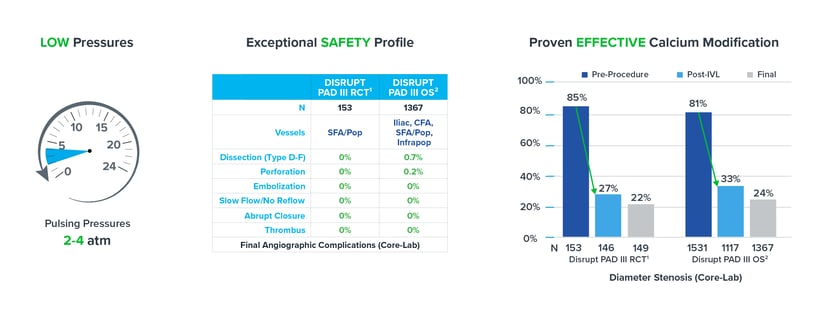
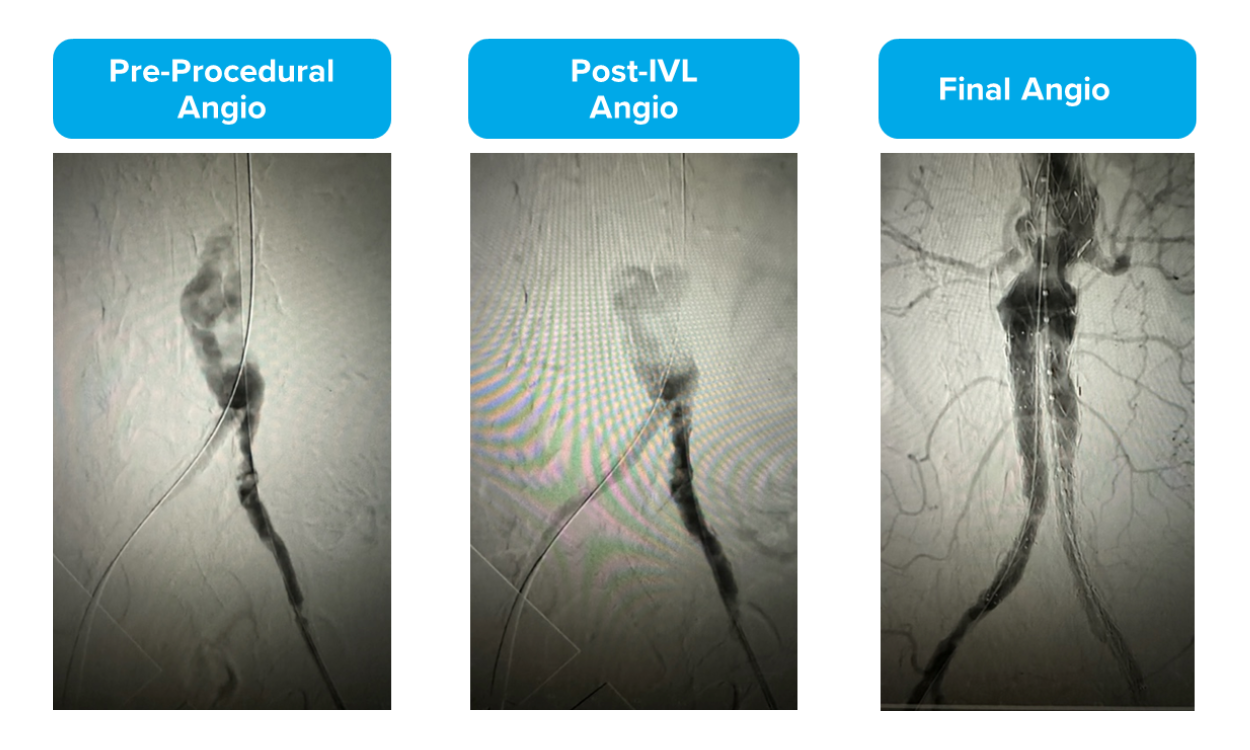
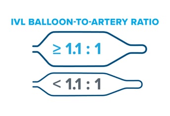
Evidence from the DISRUPT PAD clinical program (DISRUPT PAD II and DISRUPT PAD III Observational Study) shows that oversizing results in improved outcomes in terms of stenosis reduction and patency, all achieved with ultra-low treatment pressures (2-4 atm) and minimal complications, without compromising outcomes.
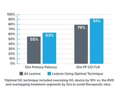
Improved Stenosis Reduction

  • The DISRUPT PAD III Observational Study of 1,373 patients represents the largest prospective ‘real-world’ evidence for the treatment of heavily calcified peripheral arterial disease.
  • Per a multivariable analysis, oversizing by 10% or greater was an independent predictor of improved stenosis reduction across multiple peripheral vessel beds but not a predictor of complications.2

Check out the DISRUPT PAD III Observational Study to learn more.

IVL Improved Patency GraphImproved Patency

  • The DISRUPT PAD II trial was a multi-center study prospectively enrolling heavily calcified, stenotic, femoropopliteal arteries with a 12-month follow-up.
  • In DISRUPT PAD II, the optimal IVL technique* (including oversizing by 10% vs. the healthy reference vessel diameter) was associated with 15% improved primary patency & rate of CD-TLR in PAD II.3
  • This technique was performed in the absence of drug, using only IVL and no drug-eluting technology.

Check out DISRUPT PAD II to learn more.

As mentioned above, Shockwave IVL works at ultra-low treatment pressures. Our data shows IVL maintains its exceptional safety profile and efficacy as a calcium modification tool even when oversized by 10%. When combined, these pillars allow you to confidently employ optimal sizing techniques in your treatment approach.

IVL Pillars Graphs

Using RVD vs. MLD for Sizing: Pre-FEVAR

By Dr. Mazin Foteh

Dr. Foteh is a paid consultant of Shockwave Medical.

As you can see, while Dr. Foteh measured the minimum lumen diameter (MLD) to be 4.5 mm, the healthy reference vessel diameter (RVD) measured to 9.5 mm. To optimize his treatment with IVL, Dr. Foteh used a 10.0 mm Shockwave L6.

Size for Success_Foteh Case Feature

Read the article, “Adding 8-12mm diameter devices to the Shockwave Peripheral Intravascular Lithotripsy toolkit,” to learn more.

Size for Success with Peripheral IVL: Interview & Case Review with Dr. Paul Foley

In this sizing discussion and case review, Dr. Paul Foley elaborates on his journey using Shockwave IVL and what led him to oversize peripheral IVL by 10% to receive optimized results.

Dr. Foley further shares his experience through reviewing three cases:

  • Case 1: Calcified Bilateral Common Iliac Artery Stenoses
  • Case 2: Calcified Left Popliteal Stenosis
  • Case 3: BTK Intervention for CLTI

Size for Success Video Series

Size for Success video series thumbnailIn this 3-part video series, Dr. Eric Secemsky (Interventional Cardiologist, Beth Israel Deaconess in Boston, MA) and Dr. Michael Siah (Vascular Surgeon, UT Southwestern Medical Center in Dallas, TX) discuss appropriate peripheral intravascular lithotripsy (IVL) sizing — the what, why and how — for optimal clinical results.

  • Part 1: Why We Undersize Our Endovascular Tools & the Importance of IVUS
  • Part 2: Peripheral IVL Sizing for Optimal Results
  • Part 3: Putting Optimal Peripheral IVL Sizing Into Action – Case Review

Check out the video series on HMP Global Learning Network.

Peer-to-Peer Articles

In the latest Endovascular Today article on Sizing for Success, Drs. Sasanka Jayasuriya and Paul J. Foley discuss:

  • How they size their endovascular tools
  • Their experience oversizing peripheral IVL by 10%
  • Their preferred imaging modalities
  • Multiple case examples highlighting their sizing approach

Now in Journal of Vascular and Interventional Radiology, Drs. Patrick Harty and Varshana Gurusamy share their experience oversizing with Peripheral IVL and share two case reviews:

  • Popliteal Disease in CLTI patient
  • Below the Knee Disease in CLTI patient

Size for Success Physician Authors


The physicians featured are paid consultants for Shockwave Medical.

1: Kereiakes et. al. J Am Coll Cardiol Intv 2021.

2: Data on file at Shockwave Medical.

3: Brodmann et al. Catheter Cardiovascular Interv. 2018; 1-8.

4: Tepe et al, J Am Coll Cardiol Intv 2021.

5: Armstrong E, VIVA Late Breaking Clinical Trial 2022.

Important Safety Information

In the United States: Rx only.

Indications for Use—The Shockwave Medical Intravascular Lithotripsy (IVL) System is intended for lithotripsy-enhanced balloon dilatation of lesions, including calcified lesions, in the peripheral vasculature, including the iliac, femoral, ilio-femoral, popliteal, infra-popliteal, and renal arteries. Not for use in the coronary, carotid or cerebral vasculature.

Contraindications—Do not use if unable to pass 0.014″ (M5, M5+, S4, E8) or 0.018″ (L6) guidewire across the lesion-Not intended for treatment of in-stent restenosis or in coronary, carotid, or cerebrovascular arteries.

Warnings—Only to be used by physicians who are familiar with interventional vascular procedures—Physicians must be trained prior to use of the device—Use the generator in accordance with recommended settings as stated in the Operator’s Manual.

Precautions—use only the recommended balloon inflation medium—Appropriate anticoagulant therapy should be administered by the physician—Decision regarding use of distal protection should be made based on physician assessment of treatment lesion morphology.

Adverse effects–Possible adverse effects consistent with standard angioplasty include–Access site complications–Allergy to contrast or blood thinner–Arterial bypass surgery—Bleeding complications—Death—Fracture of guidewire or device—Hypertension/Hypotension—Infection/sepsis—Placement of a stent—renal failure—Shock/pulmonary edema—target vessel stenosis or occlusion—Vascular complications. Risks unique to the device and its use—Allergy to catheter material(s)— Device malfunction or failure—Excess heat at target site.

Prior to use, please reference the Instructions for Use for more information on indications, contraindications, warnings, precautions and adverse events. https://discover.shockwavemedical.com/ifu

Please contact your local Shockwave representative for specific country availability.

As one of the biggest calcium publications of 2023, JACC: Cardiovascular Interventions’ inclusion of the “Direct Comparison of Rotational vs Orbital Atherectomy for Calcified Lesions Guided by Optical Coherence Tomography” study, also known as DIRO, was notable as the first prospective randomized trial comparing rotational atherectomy (RA) and orbital atherectomy (OA). In the below Q&A, we invited its lead author Dr. Naotaka Okamoto of Osaka Rosai Hospital in Osaka, Japan to share his interpretation of the findings, how the results may impact the role of RA and OA in the cath lab, and the possibility of future research on the topic.

What makes the DIRO study unique?

Dr. Okamoto: The DIRO study is the first prospective randomized trial to compare the OCT findings and clinical outcomes in patients treated with rotational atherectomy (RA) versus orbital atherectomy (OA). All procedures were performed under OCT guidance in this study. While there are several previous studies comparing OCT findings and clinical outcomes after RA and OA, they were retrospective, and not all PCIs were performed with OCT. Therefore, the “prospective” and “OCT-guided” aspects of the DIRO study are unique points.

What were the key findings of the DIRO study and what key factors do you think led to them?

Dr. Okamoto: In this study, we analyzed both pre- and post-atherectomy OCT images to evaluate accurate and pure tissue modification resulting from atherectomy devices, and we observed greater tissue modification after RA compared to OA. Interventional cardiologists familiar with IVUS or OCT might have the impression that RA can achieve larger tissue modification than OA. However, not many interventional cardiologists are familiar with intracoronary imaging. For them, the result can be surprising. Furthermore, the finding that greater stent expansion resulted in larger tissue modification after RA than OA was surprising to me.

Do the study results align with what you personally experience in clinical practice?

Dr. Okamoto: As I mentioned above, I have the impression that RA can achieve larger tissue modification than OA and the DIRO study confirmed my impression. Furthermore, the finding that a bigger burr of RA can create a bigger tissue modification aligns with my experience.

Has DIRO generated any follow-on research ideas?

Dr. Okamoto: Yes, we are considering substudies of the DIRO study. For example, exploring the risk factors of slow-flow phenomenon and relevant factors of stent under-expansion after adequate tissue modification could be interesting. Additionally, comparison OCT findings after IVL, RA and OA is also intriguing, and we are currently prospectively collecting OCT images after IVL.

Given the findings of the study, are there certain clinical situations that you would still advocate for the use of OA over RA despite its decreased tissue modification ability?

Dr. Okamoto: Rotational atherectomy (RA) and orbital atherectomy (OA) are approaches to reduce calcium volume, and the DIRO study demonstrated that RA can eliminate more calcium. RA is favorable according to this result, but the DIRO study also demonstrated that RA tended to cause slow or no flow immediately after atherectomy compared with OA. This is probably explained by 3 reasons: 1) the amount of ablated plaque was small with OA; 2) OA is not likely to lead to burr wedge during atherectomy, because of its small burr size; and 3) OA can modify only target calcified lesions, whereas RA with large burr occasionally modifies not only target calcified lesions but proximal diseased lesions, even when they contain elastic tissue. Therefore, there is a stage at which OA plays an active role, especially for patients with a low ejection fractions or the last remaining coronary artery after old myocardial infarction, in whom slow or no flow can be critical even if it is transient.


Okamoto, N., Egami, Y., Nohara, H., Kawanami, S., Sugae, H., Kawamura, A., Ukita, K., Matsuhiro, Y., Nakamura, H., Yasumoto, K., Tsuda, M., Matsunaga-Lee, Y., Yano, M., Nishino, M., & Tanouchi, J. (2023). Direct comparison of rotational vs orbital atherectomy for calcified lesions guided by optical coherence tomography. JACC: Cardiovascular Interventions, 16(17), 2125–2136. https://doi.org/10.1016/j.jcin.2023.06.016

Views expressed are those of the author and not necessarily those of Shockwave Medical.

Dr. Naotaka Okamoto is a paid consultant for Shockwave Medical. See Important Safety information below.

Coronary Important Safety Information:

In the United States: Rx only 

Indications for Use— The Shockwave Intravascular Lithotripsy (IVL) System with the Shockwave C2+ Coronary IVL Catheter is indicated for lithotripsy-enabled, low-pressure balloon dilatation of severely calcified, stenotic de novo coronary arteries prior to stenting.

Contraindications— The Shockwave C2+ Coronary IVL System is contraindicated for the following: This device is not intended for stent delivery. This device is not intended for use in carotid or cerebrovascular arteries.

Warnings—Use the IVL Generator in accordance with recommended settings as stated in the Operator’s Manual. The risk of a dissection or perforation is increased in severely calcified lesions undergoing percutaneous treatment, including IVL. Appropriate provisional interventions should be readily available. Balloon loss of pressure was associated with a numerical increase in dissection which was not statistically significant and was not associated with MACE. Analysis indicates calcium length is a predictor of dissection and balloon loss of pressure. IVL generates mechanical pulses which may cause atrial or ventricular capture in bradycardic patients. In patients with implantable pacemakers and defibrillators, the asynchronous capture may interact with the sensing capabilities. Monitoring of the electrocardiographic rhythm and continuous arterial pressure during IVL treatment is required. In the event of clinically significant hemodynamic effects, temporarily cease delivery of IVL therapy.

Precautions— Only to be used by physicians trained in angiography and intravascular coronary procedures. Use only the recommended balloon inflation medium. Hydrophilic coating to be wet only with normal saline or water and care must be taken with sharp objects to avoid damage to the hydrophilic coating. Appropriate anticoagulant therapy should be administered by the physician. Precaution should be taken when treating patients with previous stenting within 5mm of target lesion.

Potential adverse effects consistent with standard based cardiac interventions include– Abrupt vessel closure – Allergic reaction to contrast medium, anticoagulant and/or antithrombotic therapy-Aneurysm-Arrhythmia-Arteriovenous fistula-Bleeding complications-Cardiac tamponade or pericardial effusion-Cardiopulmonary arrest-Cerebrovascular accident (CVA)-Coronary artery/vessel occlusion, perforation, rupture or dissection-Coronary artery spasm-Death-Emboli (air, tissue, thrombus or atherosclerotic emboli)-Emergency or nonemergency coronary artery bypass surgery-Emergency or nonemergency percutaneous coronary intervention-Entry site complications-Fracture of the guide wire or failure/malfunction of any component of the device that may or may not lead to device embolism, dissection, serious injury or surgical intervention-Hematoma at the vascular access site(s)- Hemorrhage-Hypertension/Hypotension-Infection/sepsis/fever-Myocardial Infarction-Myocardial Ischemia or unstable angina-Pain-Peripheral Ischemia-Pseudoaneurysm-Renal failure/insufficiency-Restenosis of the treated coronary artery leading to revascularization-Shock/pulmonary edema-Slow flow, no reflow, or abrupt closure of coronary artery-Stroke Thrombus-Vessel closure, abrupt-Vessel injury requiring surgical repair-Vessel dissection, perforation, rupture, or spasm.

Risks identified as related to the device and its use: Allergic/immunologic reaction to the catheter material(s) or coating-Device malfunction, failure, or balloon loss of pressure leading to device embolism, dissection, serious injury or surgical intervention-Atrial or ventricular extra systole-Atrial or ventricular capture.

Prior to use, please reference the Instructions for Use for more information on indications, contraindications, warnings, precautions and adverse events. https://shockwavemedical.com/IFU


Dr. Nieves Gonzalo is a paid consultant of Shockwave Medical.

Important Safety Information

In the United States: Rx only

Indications for Use— The Shockwave Intravascular Lithotripsy (IVL) System with the Shockwave C2 and C2+ Coronary IVL Catheter is indicated for lithotripsy-enabled, low-pressure balloon dilatation of severely calcified, stenotic de novo coronary arteries prior to stenting.

Contraindications— The Shockwave C2 and C2+ Coronary IVL System is contraindicated for the following: This device is not intended for stent delivery. This device is not intended for use in carotid or cerebrovascular arteries.

Warnings—Use the IVL Generator in accordance with recommended settings as stated in the Operator’s Manual. The risk of a dissection or perforation is increased in severely calcified lesions undergoing percutaneous treatment, including IVL. Appropriate provisional interventions should be readily available. Balloon loss of pressure was associated with a numerical increase in dissection which was not statistically significant and was not associated with MACE. Analysis indicates calcium length is a predictor of dissection and balloon loss of pressure. IVL generates mechanical pulses which may cause atrial or ventricular capture in bradycardic patients. In patients with implantable pacemakers and defibrillators, the asynchronous capture may interact with the sensing capabilities. Monitoring of the electrocardiographic rhythm and continuous arterial pressure during IVL treatment is required. In the event of clinically significant hemodynamic effects, temporarily cease delivery of IVL therapy.

Precautions— Only to be used by physicians trained in angiography and intravascular coronary procedures. Use only the recommended balloon inflation medium. Hydrophilic coating to be wet only with normal saline or water and care must be taken with sharp objects to avoid damage to the hydrophilic coating. Appropriate anticoagulant therapy should be administered by the physician. Precaution should be taken when treating patients with previous stenting within 5mm of target lesion.

Potential adverse effects consistent with standard based cardiac interventions include– Abrupt vessel closure – Allergic reaction to contrast medium, anticoagulant and/or antithrombotic therapy-Aneurysm-Arrhythmia-Arteriovenous fistula-Bleeding complications-Cardiac tamponade or pericardial effusion-Cardiopulmonary arrest-Cerebrovascular accident (CVA)-Coronary artery/vessel occlusion, perforation, rupture or dissection-Coronary artery spasm-Death-Emboli (air, tissue, thrombus or atherosclerotic emboli)-Emergency or nonemergency coronary artery bypass surgery-Emergency or nonemergency percutaneous coronary intervention-Entry site complications-Fracture of the guide wire or failure/malfunction of any component of the device that may or may not lead to device embolism, dissection, serious injury or surgical intervention-Hematoma at the vascular access site(s)- Hemorrhage-Hypertension/Hypotension-Infection/sepsis/fever-Myocardial Infarction-Myocardial Ischemia or unstable angina-Pain-Peripheral Ischemia-Pseudoaneurysm-Renal failure/insufficiency-Restenosis of the treated coronary artery leading to revascularization-Shock/pulmonary edema-Slow flow, no reflow, or abrupt closure of coronary artery-Stroke Thrombus-Vessel closure, abrupt-Vessel injury requiring surgical repair-Vessel dissection, perforation, rupture, or spasm.

Risks identified as related to the device and its use: Allergic/immunologic reaction to the catheter material(s) or coating-Device malfunction, failure, or balloon loss of pressure leading to device embolism, dissection, serious injury or surgical intervention-Atrial or ventricular extrasystole-Atrial or ventricular capture.

Prior to use, please reference the Instructions for Use for more information on warnings, precautions and adverse events. www.shockwavemedical.com/IFU.

In this Cath Lab Digest article, Dr. Brian Jefferson of Centennial Heart, HCA Tristar Centennial Medical Center, Nashville, Tennessee explains why the 40 additional pulses in the Shockwave C2+ catheter allows for an “IVL-first” strategy in lesions he previously reserved for atherectomy (i.e. long, diffuse lesions and multi-vessel disease).

This article was developed in collaboration with Cath Lab Digest to bring you the latest calcium insights from the U.S. coronary interventional experts who know it best. The Calcium Corner, contains a series of articles where operators share their perspective on the challenges and treatment of coronary artery calcium. Read about experts’ clinical experience, case studies and treatment algorithms with Coronary IVL – hope you enjoy the content.


Dr. Brian Jefferson is a paid consultant for Shockwave Medical.

Coronary Important Safety Information:

In the United States: Rx only.

Indications for Use—The Shockwave Intravascular Lithotripsy (IVL) System with the Shockwave C2 Coronary IVL Catheter is indicated for lithotripsy-enabled, low-pressure balloon dilatation of severely calcified, stenotic de novo coronary arteries prior to stenting.

Contraindications—The Shockwave C2 Coronary IVL System is contraindicated for the following: This device is not intended for stent delivery. This device is not intended for use in carotid or cerebrovascular arteries.

Warnings— Use the IVL Generator in accordance with recommended settings as stated in the Operator’s Manual. The risk of a dissection or perforation is increased in severely calcified lesions undergoing percutaneous treatment, including IVL. Appropriate provisional interventions should be readily available. Balloon loss of pressure was associated with a numerical increase in dissection which was not statistically significant and was not associated with MACE.  Analysis indicates calcium length is a predictor of dissection and balloon loss of pressure.  IVL generates mechanical pulses which may cause atrial or ventricular capture in bradycardic patients. In patients with implantable pacemakers and defibrillators, the asynchronous capture may interact with the sensing capabilities. Monitoring of the electrocardiographic rhythm and continuous arterial pressure during IVL treatment is required.  In the event of clinically significant hemodynamic effects, temporarily cease delivery of IVL therapy.

Precautions— Only to be used by physicians trained in angiography and intravascular coronary procedures. Use only the recommended balloon inflation medium. Hydrophilic coating to be wet only with normal saline or water and care must be taken with sharp objects to avoid damage to the hydrophilic coating. Appropriate anticoagulant therapy should be administered by the physician. Precaution should be taken when treating patients with previous stenting within 5mm of target lesion.

Potential adverse effects consistent with standard based cardiac interventions include– Abrupt vessel closure – Allergic reaction to contrast medium, anticoagulant and/or antithrombotic therapy-Aneurysm-Arrhythmia-Arteriovenous fistula-Bleeding complications-Cardiac tamponade or pericardial effusion-Cardiopulmonary arrest-Cerebrovascular accident (CVA)-Coronary artery/vessel occlusion, perforation, rupture or dissection-Coronary artery spasm-Death-Emboli (air, tissue, thrombus or atherosclerotic emboli)-Emergency or non-emergency coronary artery bypass surgery-Emergency or non-emergency percutaneous coronary intervention-Entry site complications-Fracture of the guide wire or failure/malfunction of any component of the device that may or may not lead to device embolism, dissection, serious injury or surgical intervention-Hematoma at the vascular access site(s)-Hemorrhage-Hypertension/Hypotension-Infection/sepsis/fever-Myocardial Infarction-Myocardial Ischemia or unstable angina-Pain-Peripheral Ischemia-Pseudoaneurysm-Renal failure/insufficiency-Restenosis of the treated coronary artery leading to revascularization-Shock/pulmonary edema-Slow flow, no reflow, or abrupt closure of coronary artery-Stroke-Thrombus-Vessel closure, abrupt-Vessel injury requiring surgical repair-Vessel dissection, perforation, rupture, or spasm. Risks identified as related to the device and its use: Allergic/immunologic reaction to the catheter material(s) or coating-Device malfunction, failure, or balloon loss of pressure leading to device embolism, dissection, serious injury or surgical intervention-Atrial or ventricular extrasystole-Atrial or ventricular capture.

Prior to use, please reference the Instructions for Use for more information on warnings, precautions and adverse events.  https://shockwavemedical.com/IFU

Please contact your local Shockwave representative for specific country availability and refer to the Shockwave C2 instructions for use containing important safety information.

A new publication titled “Use of Calcium Modification during Percutaneous Coronary Intervention after Introduction of Coronary Intravascular Lithotripsy” details an overall increase in the use of calcium modification tools for percutaneous coronary interventions (PCI) and a rapid uptake in the use of coronary IVL since the U.S. launch of Shockwave C2 in early 2021. The research, led by Dr. Neel Butala, Assistant Professor, Medicine-Cardiology at the University of Colorado School of Medicine, was recently published in the Journal of the Society for Cardiovascular Angiography and Interventions (JSCAI).

The research analyzed data from the ACC/NCDR® (National Cardiovascular Data Registry) CathPCI® Registry from April 2018 to December 2022 and revealed that in less than two years, coronary IVL expanded the appreciation of lesion preparation and use of calcium modification tools more broadly and has rapidly become the most used calcium modification technology in the U.S. Of the PCI cases analyzed during the second quarter of 2018, 8.7% were performed with calcium modification tools, including cutting or scoring balloon angioplasty, rotational and orbital atherectomy. By the fourth quarter of 2020, this number increased to 11.1%. By the fourth quarter of 2022, in conjunction with the U.S. commercial introduction of coronary IVL in early 2021, the use of calcium modification tools increased to 16.0% of PCI cases. In only 18 months of availability in the U.S., coronary IVL was used in 7.8% of PCI cases, while the use of atherectomy decreased from 5.4% to 4.4% of cases during the same timeframe.  

“Our research shows a rapid uptake in coronary IVL, likely driven by its safety profile and ease of use relative to atherectomy, which makes calcium modification as simple as balloon angioplasty,” said Dr. Butala, the first author on the publication. “I would like to thank SCAI for the opportunity to conduct this research as part of the society’s Early Career Research Grants program and I look forward conducting deeper analyses into the ACC/NCDR CathPCI Registry, particularly with respect to identifying predictors of calcium modification strategies and their correlated outcomes.”

Read the Publication


Dr. Neel Butala is a paid consultant for Shockwave Medical.

Coronary Important Safety Information:

In the United States: Rx only.

Indications for Use—The Shockwave Intravascular Lithotripsy (IVL) System with the Shockwave C2 Coronary IVL Catheter is indicated for lithotripsy-enabled, low-pressure balloon dilatation of severely calcified, stenotic de novo coronary arteries prior to stenting.

Contraindications—The Shockwave C2 Coronary IVL System is contraindicated for the following: This device is not intended for stent delivery. This device is not intended for use in carotid or cerebrovascular arteries.

Warnings— Use the IVL Generator in accordance with recommended settings as stated in the Operator’s Manual. The risk of a dissection or perforation is increased in severely calcified lesions undergoing percutaneous treatment, including IVL. Appropriate provisional interventions should be readily available. Balloon loss of pressure was associated with a numerical increase in dissection which was not statistically significant and was not associated with MACE.  Analysis indicates calcium length is a predictor of dissection and balloon loss of pressure.  IVL generates mechanical pulses which may cause atrial or ventricular capture in bradycardic patients. In patients with implantable pacemakers and defibrillators, the asynchronous capture may interact with the sensing capabilities. Monitoring of the electrocardiographic rhythm and continuous arterial pressure during IVL treatment is required.  In the event of clinically significant hemodynamic effects, temporarily cease delivery of IVL therapy.

Precautions— Only to be used by physicians trained in angiography and intravascular coronary procedures. Use only the recommended balloon inflation medium. Hydrophilic coating to be wet only with normal saline or water and care must be taken with sharp objects to avoid damage to the hydrophilic coating. Appropriate anticoagulant therapy should be administered by the physician. Precaution should be taken when treating patients with previous stenting within 5mm of target lesion.

Potential adverse effects consistent with standard based cardiac interventions include– Abrupt vessel closure – Allergic reaction to contrast medium, anticoagulant and/or antithrombotic therapy-Aneurysm-Arrhythmia-Arteriovenous fistula-Bleeding complications-Cardiac tamponade or pericardial effusion-Cardiopulmonary arrest-Cerebrovascular accident (CVA)-Coronary artery/vessel occlusion, perforation, rupture or dissection-Coronary artery spasm-Death-Emboli (air, tissue, thrombus or atherosclerotic emboli)-Emergency or non-emergency coronary artery bypass surgery-Emergency or non-emergency percutaneous coronary intervention-Entry site complications-Fracture of the guide wire or failure/malfunction of any component of the device that may or may not lead to device embolism, dissection, serious injury or surgical intervention-Hematoma at the vascular access site(s)-Hemorrhage-Hypertension/Hypotension-Infection/sepsis/fever-Myocardial Infarction-Myocardial Ischemia or unstable angina-Pain-Peripheral Ischemia-Pseudoaneurysm-Renal failure/insufficiency-Restenosis of the treated coronary artery leading to revascularization-Shock/pulmonary edema-Slow flow, no reflow, or abrupt closure of coronary artery-Stroke-Thrombus-Vessel closure, abrupt-Vessel injury requiring surgical repair-Vessel dissection, perforation, rupture, or spasm. Risks identified as related to the device and its use: Allergic/immunologic reaction to the catheter material(s) or coating-Device malfunction, failure, or balloon loss of pressure leading to device embolism, dissection, serious injury or surgical intervention-Atrial or ventricular extrasystole-Atrial or ventricular capture.

Prior to use, please reference the Instructions for Use for more information on warnings, precautions and adverse events.  https://shockwavemedical.com/IFU

Please contact your local Shockwave representative for specific country availability and refer to the Shockwave C2 instructions for use containing important safety information.

Calcium Masterclass Class 11, featuring Professor James C. Spratt, Dr. Robert Riley, Dr. Ziad Ali, Dr. Jonathan Hill, Dr. Margaret McEntegart and Dr. Claudia Cosgrove as they discuss nodular calcium and a live case review at St. George’s Hospital in London.

Calcium Masterclass Class 12, featuring Dr. Ziad Ali, Dr. Margaret McEntegart, Professor James C. Spratt, Dr. Jonathan Hill, Dr. Robert Riley and Dr. Simon Wilson as they explore eccentric calcium in a live case at St. George’s Hospital in London.


The physicians featured are paid consultants for Shockwave Medical.