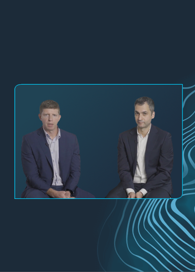
IVL in Real-World European Population: Q&A with Dr. Sandeep Basavarajaiah & Dr. Alfonso Ielasi
Dr. Sandeep Basavarajaiah, consultant cardiologist at Heartlands Hospital in Birmingham, UK, and other leading UK and Italian cardiologists published their initial real-world experience with Intravascular Lithotripsy (IVL) in a real-world population, in Catheter Cardiovasc Interv: “Intravascular lithotripsy in calcified-coronary lesions: A real-world observational, European multicenter study”.
We had the privilege of connecting with Dr. Sandeep Basavarajaiah and with Dr. Alfonso Ielasi, from Milan, Italy, to learn more about the publication and its key takeaways.
Read the publication and enjoy their Q&A:
What does this study add to interventionalists’ daily practice?
Dr. Basavarajaiah: This study provides the safety data on IVL use in real-world population from six-high volume centres that undertake complex coronary intervention. In addition, it also demonstrates the high success rate in completing the procedure with very low event rates during this short-term follow-up.
How will you describe the baseline demographics of patients included in this study and how does it compare with DISRUPT CAD trials already published?
Dr. Ielasi: From a clinical point of view our study reflects a different nature compared to the DISRUPT CAD studies. Typical clinical predictors of events, such diabetes and CKD, were more represented in our “all-comers” registry as well as the more aggressive interventional approaches, e.g. “rotatripsy”. The lack of a systematic use of intracoronary imaging in our study reflects a real-world practice according to country and regional reimbursement. However, this aspect did not influence the clinical outcome.
From your perspective what are the key takeaways from this publication?
Dr. Basavarajaiah: Our study from a real-world practice has shown that the use of IVL in complex calcified coronary lesions appears safe with low rates of complications and high rates of procedural success. In addition, the clinical outcomes are promising with low even rates. This should encourage operators to embrace this novel technology in their daily clinical practice.
Intravascular imaging was only used in ~1/4 of the cases. What drives the need of use intravascular imaging in this group of patients? Do you think that using intracoronary imaging is mandatory in a real-world setting in order to attain good results with IVL?
Dr. Ielasi: In our study, the use of intravascular imaging was left to operator’s discretion and it was mostly performed in the initial IVL experience (led by the curiosity to assess the IVL effect on calcified lesions) and in case of intra-stent lesions (off label use). Although intravascular imaging is of paramount importance to appreciate the type and extension of the calcifications in the vessel wall, in our experience not using intracoronary imaging to guide PCI did not result in poorer clinical outcomes.
When intravascular imaging isn’t available how do you decide to use IVL based on angio alone?
Dr. Basavarajaiah: Degree of calcium can be analysed on the angiogram, although not as accurately as from intra-vascular imaging. If intra-vascular imaging is not available, we would suggest considering IVL only if conventional devices failed to expand the lesion (bailout use of IVL).
In this paper you present a very simplified algorithm. With the current algorithms previously developed, what compelled you to develop this straightforward and imaging-free algorithm?
Dr. Ielasi: This algorithm provides a simple and practical decision making approach, to treat resistant coronary lesions without the need of intracoronary imaging, while providing some suggestion on when IVL usage should be considered.
Dr. Sandeep Basavarajaiah and Dr. Alfonso Ielasi are paid consultants of Shockwave Medical.
Coronary Important Safety Information:
In the United States: Rx only.
Indications for Use—The Shockwave Intravascular Lithotripsy (IVL) System with the Shockwave C2 Coronary IVL Catheter is indicated for lithotripsy-enabled, low-pressure balloon dilatation of severely calcified, stenotic de novo coronary arteries prior to stenting.
Contraindications—The Shockwave C2 Coronary IVL System is contraindicated for the following: This device is not intended for stent delivery. This device is not intended for use in carotid or cerebrovascular arteries.
Warnings— Use the IVL Generator in accordance with recommended settings as stated in the Operator’s Manual. The risk of a dissection or perforation is increased in severely calcified lesions undergoing percutaneous treatment, including IVL. Appropriate provisional interventions should be readily available. Balloon loss of pressure was associated with a numerical increase in dissection which was not statistically significant and was not associated with MACE. Analysis indicates calcium length is a predictor of dissection and balloon loss of pressure. IVL generates mechanical pulses which may cause atrial or ventricular capture in bradycardic patients. In patients with implantable pacemakers and defibrillators, the asynchronous capture may interact with the sensing capabilities. Monitoring of the electrocardiographic rhythm and continuous arterial pressure during IVL treatment is required. In the event of clinically significant hemodynamic effects, temporarily cease delivery of IVL therapy.
Precautions— Only to be used by physicians trained in angiography and intravascular coronary procedures. Use only the recommended balloon inflation medium. Hydrophilic coating to be wet only with normal saline or water and care must be taken with sharp objects to avoid damage to the hydrophilic coating. Appropriate anticoagulant therapy should be administered by the physician. Precaution should be taken when treating patients with previous stenting within 5mm of target lesion.
Potential adverse effects consistent with standard based cardiac interventions include– Abrupt vessel closure – Allergic reaction to contrast medium, anticoagulant and/or antithrombotic therapy-Aneurysm-Arrhythmia-Arteriovenous fistula-Bleeding complications-Cardiac tamponade or pericardial effusion-Cardiopulmonary arrest-Cerebrovascular accident (CVA)-Coronary artery/vessel occlusion, perforation, rupture or dissection-Coronary artery spasm-Death-Emboli (air, tissue, thrombus or atherosclerotic emboli)-Emergency or non-emergency coronary artery bypass surgery-Emergency or non-emergency percutaneous coronary intervention-Entry site complications-Fracture of the guide wire or failure/malfunction of any component of the device that may or may not lead to device embolism, dissection, serious injury or surgical intervention-Hematoma at the vascular access site(s)-Hemorrhage-Hypertension/Hypotension-Infection/sepsis/fever-Myocardial Infarction-Myocardial Ischemia or unstable angina-Pain-Peripheral Ischemia-Pseudoaneurysm-Renal failure/insufficiency-Restenosis of the treated coronary artery leading to revascularization-Shock/pulmonary edema-Slow flow, no reflow, or abrupt closure of coronary artery-Stroke-Thrombus-Vessel closure, abrupt-Vessel injury requiring surgical repair-Vessel dissection, perforation, rupture, or spasm. Risks identified as related to the device and its use: Allergic/immunologic reaction to the catheter material(s) or coating-Device malfunction, failure, or balloon loss of pressure leading to device embolism, dissection, serious injury or surgical intervention-Atrial or ventricular extrasystole-Atrial or ventricular capture.
Prior to use, please reference the Instructions for Use for more information on warnings, precautions and adverse events. https://shockwavemedical.com/IFU
Please contact your local Shockwave representative for specific country availability and refer to the Shockwave C2 instructions for use containing important safety information.