This recorded webinar features an in‑depth, case‑based discussion focused on the treatment of heavily calcified below‑the‑knee (BTK) disease in patients with peripheral artery disease (PAD) and chronic limb‑threatening ischemia (CLTI). The session is moderated by Misty Humphries, MD, MAS, vascular surgeon at UC Davis Medical Center in Sacramento, CA, and includes expert faculty Venita Chandra, MD of Stanford University (Stanford, CA) and Leigh Ann O’Banion, MD of UCSF Fresno (Fresno, CA).

Dr. Humphries opens the webinar with an overview of the paradigm shift in BTK calcium management, highlighting the clinical challenges posed by diffuse, medial calcification, vessel recoil, and small vessel diameter in CLTI patients. She reviews best practices for intravascular lithotripsy (IVL), emphasizing the importance of intravascular ultrasound (IVUS)‑guided sizing, appropriate balloon oversizing relative to reference vessel diameter, and low‑pressure inflation to optimize calcium fracture and luminal gain while maintaining a strong safety profile. Technical features and use cases for Shockwave E8 and the Javelin forward‑emitting IVL catheter are also discussed.

Dr. Chandra reviews outcomes from the FORWARD study and new 12‑month data from the DISRUPT BTK II trial. She highlights the real‑world nature of these studies, which include patients with severe calcification, chronic total occlusions, diabetes, and end‑stage renal disease. Key findings discussed include low angiographic complication rates, meaningful reductions in diameter stenosis, favorable patency and freedom‑from‑TLR outcomes, high freedom from major amputation, and improvements in Rutherford classification, wound healing, and quality‑of‑life metrics.

Dr. O’Banion concludes the program by presenting real‑world cases that illustrate access planning, lesion crossing strategies, and treatment of diffuse tibial and pedal disease. Her cases demonstrate practical use of IVL technologies to modify calcified vessels, restore inline flow to wound‑related angiosomes, and achieve limb salvage without the need for stenting or surgical bypass in selected patients.

Throughout the discussion, the faculty emphasize multidisciplinary limb‑salvage decision‑making, thoughtful device selection, and the evolving role of IVL in treating complex BTK disease.

Learn more about DISRUPT BTK II here.


Drs. Humphries, Chandra, and O’Banion are paid consultants for Shockwave Medical.

Shockwave IVL: In the U.S.: Rx only. Prior to use, please reference the Important Safety Information for more information on indications, contraindications, warnings, precautions and adverse events.
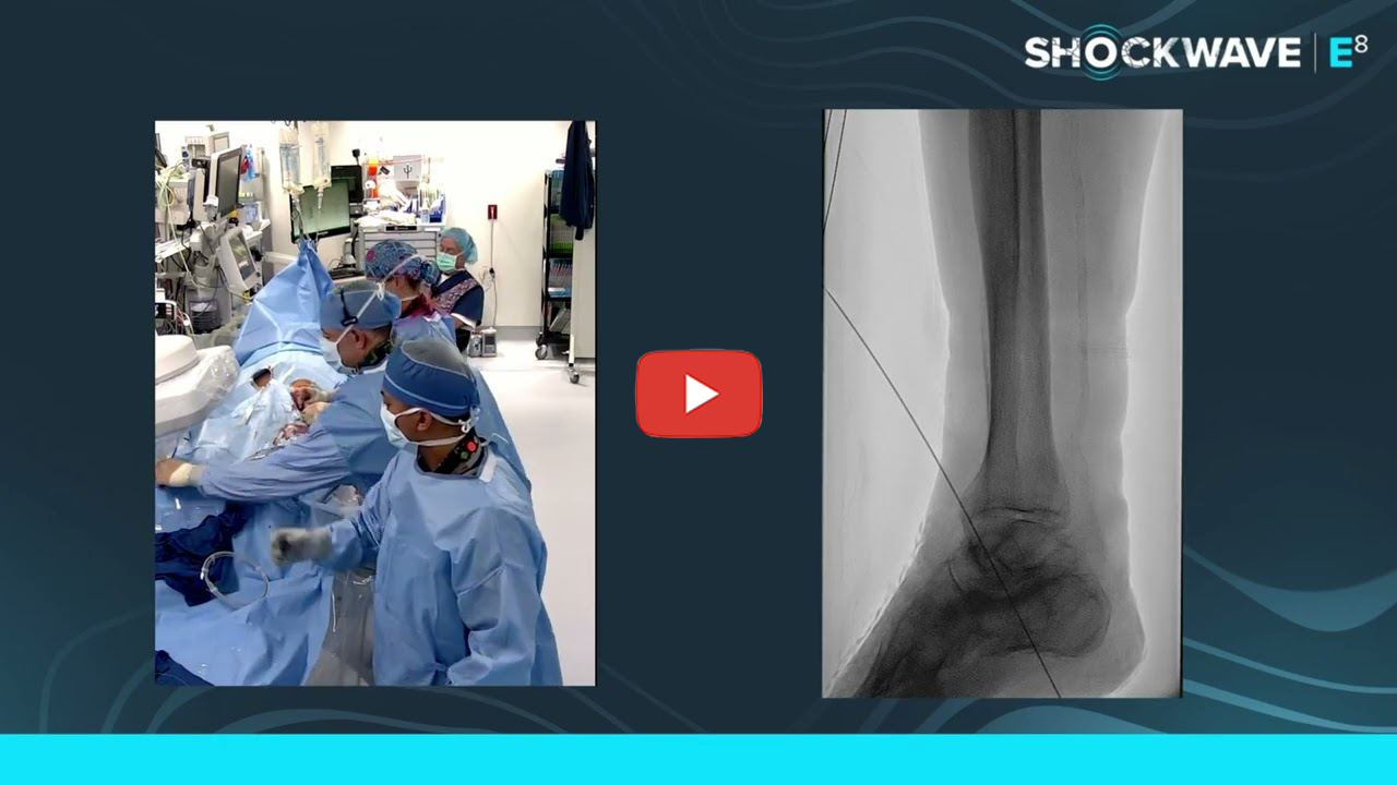
Dr. Foteh shares an IVL procedure on an 85 year-old female with history of coronary artery disease (CAD), hypertension, hyperlipidemia, diabetes, who presented with a calcaneal ulcer and abnormal ankle-brachial and toe-brachial indices.

After successfully crossing the lesion, Dr. Foteh advances a Shockwave IVL E8 5.0 mm x 80 mm catheter delivering all pulses. IVL therapy is followed by a 5.0 mm x 40 mm drug coated balloon (DCB) resulting in improved flow with no dissection or embolic complications.


Dr. Foteh is a paid consultant of Shockwave Medical.

Shockwave IVL: In the U.S.: Rx only. Prior to use, please reference the Important Safety Information for more information on indications, contraindications, warnings, precautions and adverse events.
Dr. Foteh presents a 79 year-old male patient with history of coronary artery disease (CAD), hypertension, hyperlipidemia, peripheral artery disease (PAD), and abnormal ankle-brachial (ABI) and toe-brachial indices (TBI).

Using a contralateral approach, Dr. Foteh successfully crosses the lesion advancing a 4.0 mm x 80 mm Shockwave IVL E8, followed by a 4.0 mm drug coated balloon (DCB) in the anterior tibial and a larger DCB in the superficial femoral artery (SFA), providing improved flow with no dissection or embolic complications.


Dr. Foteh is a paid consultant of Shockwave Medical.

Shockwave IVL: In the U.S.: Rx only. Prior to use, please reference the Important Safety Information for more information on indications, contraindications, warnings, precautions and adverse events.
Dr. Foley presents an 88 year-old male with a history of heart failure, and non-compressible ankle-brachial index (ABI), and very reduced toe-brachial index (TBI), with a gangrenous ulcer of the right hallux.

Using an antegrade right common femoral approach, Dr. Foley successfully crosses the lesion and advances the Shockwave IVL Javelin, followed by post dilation with a 3.0 mm x 22 mm PTA balloon resulting in improved distal flow with no dissection or embolic complications.


Dr. Foley is a paid consultant of Shockwave Medical.

Shockwave IVL: In the U.S.: Rx only. Prior to use, please reference the Important Safety Information for more information on indications, contraindications, warnings, precautions and adverse events.
In this video, Dr. Venita Chandra shares 12-month outcomes from the DISRUPT BTK II trial, a prospective study evaluating IVL in a cohort of 250 patients, 200 of which have chronic limb-threatening ischemia (CLTI)—a population that is often excluded from clinical studies due to disease complexity.

The presentation highlights key clinical findings, including freedom from low intervention rates, low major amputation rates, and meaningful improvements in quality of life and wound healing. These outcomes demonstrate durable results at 12 months in severely calcified, below-the-knee disease.

Together, the data shown in this video support IVL as a frontline calcium-modification strategy* and an important part of the treatment algorithm for CLTI, reinforcing its role in improving patient outcomes and reducing amputation risk.

Watch now to learn more about how IVL is impacting outcomes in patients with CLTI.


Dr. Venita Chandra is a paid consultant of Shockwave Medical.

*Frontline Strategy: The approach of utilizing IVL as the primary treatment method, implemented without the use of atherectomy. This strategy is backed by the BTK II data based on the percentage of procedures involving pre-dilatation and post-dilatation, as well as the proportion of stenting performed. Our BTK II data indicates a notably low usage of both pre-dilatation and post-stenting in these cases.

Shockwave IVL: In the U.S.: Rx only. Prior to use, please reference the Important Safety Information for more information on indications, contraindications, warnings, precautions and adverse events.

In this video, Dr. Michael Siah from UT Southwestern in Dallas presents a complex limb-salvage case involving an 84-year-old patient with critical limb-threatening ischemia and peripheral arterial disease. The patient had significant comorbidities, including end-stage renal disease, heart failure, lupus, and a non-healing right foot ulcer, along with prior deep vein arterialization on the contralateral limb. Imaging revealed severe disease spanning the superficial femoral artery, tibial vessels, and below-the-ankle circulation, including a heavily calcified dorsalis pedis chronic total occlusion that proved difficult to treat with standard endovascular tools.

Dr. Siah walks through his strategy, detailing challenges with access, device delivery, and lesion crossing despite balloon angioplasty and intravascular lithotripsy (IVL). When conventional low-profile balloons and orbital atherectomy were unsuccessful due to delivery limitations, he turned to the Shockwave Javelin catheter where its use significantly modified the plaque and vessel compliance. This effect allowed a previously unsuccessful balloon to cross and treat the lesion, ultimately restoring inline flow to the forefoot.

The successful outcome—near-complete wound healing at six weeks and preservation of a functional transmetatarsal amputation—underscores the importance of patience, adaptability, and thoughtful tool selection when treating advanced limb ischemia.


Dr. Siah is a paid consultant of Shockwave Medical. Views expressed are those of the presenters and not necessarily those of Shockwave IVL.

Shockwave IVL: In the U.S.: Rx only. Prior to use, please reference the Important Safety Information for more information on indications, contraindications, warnings, precautions and adverse events. Please contact your local Shockwave representative for specific country availability.

Join Dr. Ziad Ali, Dr. Jai Khatri, Dr. Aloke Finn and Professor James Spratt as they discuss coronary artery calcification and the importance of understanding calcium morphologies in guiding treatment during percutaneous coronary intervention (PCI).

During the webinar, the Faculty review data from a recent cadaver-based comparative study that evaluated Intravascular Lithotripsy (IVL) alongside cutting balloons and ultra–high-pressure balloons using micro-CT, OCT, and histology. The study included calcium modification data from key calcium types—concentric, eccentric, and nodular calcium —and they explain why accurate assessment of calcium morphology is essential for selecting safe and effective treatment options. Findings from the study demonstrated that IVL consistently produces calcium fractures in most lesions, while minimizing vessel wall injury, whereas high-pressure balloons frequently damaged the vessel wall with less consistent fracture. An important consideration given the association between medial injury and adverse long-term outcomes.

Additionally, the Faculty also discuss Shockwave IVL’s mechanism of action, highlighting how ultrasonic pressure waves selectively fracture calcium based on acoustic impedance while preserving soft tissue. Clinical case examples illustrate the role of IVL in complex and high-risk lesions where procedural safety is critical, reinforcing IVL’s value as a low-risk and effective approach to calcium modification across a range of coronary anatomies.


Drs. Ali, Khatri, and Finn, and Professor Spratt are paid consultants of Shockwave Medical. Views expressed are those of the presenters and not necessarily those of Shockwave IVL.

Shockwave IVL: In the U.S.: Rx only. Prior to use, please reference the Important Safety Information for more information on indications, contraindications, warnings, precautions and adverse events. Please contact your local Shockwave representative for specific country availability.

Watch Dr. Paul Foley and Dr. Mazin Foteh for a virtual case review, moderated by Dr. Leigh Ann O’Banion, as they discuss two of their challenging below-the-knee (BTK) cases using Shockwave Intravascular Lithotripsy (IVL). The webinar showcases detailed case reviews, including the use of innovative technology such as Shockwave Javelin, highlighting its role in modifying challenging calcified lesions.

In this session, Dr. Foley presents a compelling case of an elderly male with limb-threatening ischemia, non-healing ulcers, and extensive calcified arterial disease. The discussion covers preoperative planning, including the importance of duplex ultrasound, CTA imaging, and strategic access approaches—antegrade versus pedal access—to optimize procedural success. The team emphasizes the critical role of intravascular ultrasound (IVUS) in accurately sizing vessels and characterizing plaque, which guides effective treatment choices.

Throughout the webinar, the panel explores various crossing techniques, wire escalation strategies, and the nuances of delivering energy with devices like Shockwave Javelin. They compare different atherectomy and lithotripsy tools, debating the merits of focal versus circumferential calcium modification, and discuss how these technologies can improve vessel compliance and blood flow. The importance of multidisciplinary collaboration, including insights from interventional cardiology and radiology, is also underscored as vital to advancing limb salvage efforts.


Drs. O’Banion, Foley, and Foteh are paid consultants of Shockwave Medical. Views expressed are those of the presenters and not necessarily those of Shockwave IVL.

Shockwave IVL: In the U.S.: Rx only. Prior to use, please reference the Important Safety Information for more information on indications, contraindications, warnings, precautions and adverse events. Please contact your local Shockwave representative for specific country availability.

A recently published cadaveric study compares the frequency and patterns of calcium fracture and medial injury based on the mechanism of action (MOA) of advanced calcified lesion preparation strategies.

17 cadaveric calcified lesions were randomized to one of three treatment arms of Intravascular Lithotripsy (IVL; Shockwave C2+), cutting balloons (CB; Wolverine™ Boston Scientific) and ultra-high pressure balloons (UHB; OPN NC SIS Medical). Frequency of calcium fracture and presence of medial injury – which potentially negatively affects long-term clinical outcomes due to increased neointimal thickness, higher inflammatory cell count and in-stent restenosis1,2 – were identified using gold-standard techniques of micro-CT and histology.

Shockwave C2+ produced the most calcium fracture with the least amount of medial injury by lesion and across concentric and eccentric calcium arcs. Within histological sections with concentric calcium, fractures produced by Wolverine™ and OPN NC were mostly associated with medial injury. No calcium fracture was identified for UHB within eccentric calcium.

Learn more by downloading the one-page clinical summary and viewing the video below for a roundtable discussion between the study’s principal investigators, Drs. Ziad Ali and Aloke Finn, moderated by Shockwave Medical Chief Medical Officer, Dr. Nick West.


Download Clinical Summary


Sekimoto, T et al. Comparison of Vascular Injury From Intravascular Lithotripsy, Cutting, or Ultra-High-Pressure Balloons During Coronary Calcium Modification. J Am Coll Cardiol Intv. 2025 Sep, 18 (17) 2093-2104. Cadaveric study. May not be indicative of actual clinical use.

1: Farb A, et al., Pathology of acute and chronic coronary stenting in humans. Circulation. 1999 Jan 5-12;99(1):44-52.
2: Schwartz RS, et al., Restenosis and the proportional neointimal response to coronary artery injury: results in a porcine model. J Am Coll Cardiol. 1992 Feb;19(2):267-74.

Drs. Ziad Ali and Aloke Finn are paid consultants of Shockwave Medical. Views expressed are those of the presenters and not necessarily those of Shockwave IVL.

Shockwave IVL: In the U.S.: Rx only. Prior to use, please reference the Important Safety Information for more information on indications, contraindications, warnings, precautions and adverse events. Please contact your local Shockwave representative for specific country availability.

In this informative and in-depth session, renowned vascular specialists and interventionalists come together to explore advanced strategies for managing critical limb-threatening ischemia (CLTI) and complex tibial and pedal artery disease. Moderated by Dr. John Rollo, the discussion features expert contributions from Dr. Misty Humphreys (UC Davis), Dr. Constantino Pena (Baptist Miami), and Dr. John Corl (Cincinnati), who share their insights on techniques for challenging cases involving heavily calcified vessels and non-healing foot ulcers.

Cutting-Edge Treatment Approaches

Dr. Humphreys highlights her use of the latest balloon technology, including Shockwave E8, which facilitates treating entire tibial segments with long, subnominal inflations—often exceeding three minutes—aimed at optimizing vessel compliance. She emphasizes the importance of thorough pre-procedure planning, including sizing and physiologic assessments like pedal acceleration time, toe pressures, and transcutaneous oxygen measurements, to guide treatment decisions and improve healing outcomes.

Case-Specific Strategies

The discussion delves into case-specific strategies, particularly focusing on targeting the most promising arteries, such as the anterior tibial artery, and considering collateral flow and angiosome principles to maximize tissue perfusion. Panelists discuss when to treat multiple vessels versus a more conservative approach, stressing that treatment should be tailored based on physiologic data, wound status, and patient risk.

Shockwave Javelin

Challenging cases are examined, especially those with heavily calcified lesions where crossing may be difficult. The panel introduces Shockwave Javelin, a calcium modification tool that uses forward-shifted ultrasound energy to crack calcified plaque and facilitate successful balloon crossing. They share practical tips on crossing strategies, sheath and wire selection, and the benefits of retrograde access when antegrade approaches fail.


Dr. John Rollo, Dr. John Corl, Dr. Misty Humphries, and Dr. Constantino Pena are paid consultants of Shockwave Medical. The thoughts and views expressed are of their own opinions, and do not necessarily represent those of Shockwave Medical.

Shockwave IVL: In the U.S.: Rx only. Prior to use, please reference the Important Safety Information for more information on indications, contraindications, warnings, precautions and adverse events.

Please contact your local Shockwave representative for specific country availability.