DISRUPT CAD II

By The Numbers

120
Patients At 15 Sites
94%
Severe Ca++
100%
IVL & Stent Delivery
0%
Major Dissections
0%
Perforations
0%
Slow/No Reflow

DISRUPT CAD II Study Leadership

jean fajadet
Prof. Jean Fajadet
Co-Principal Investigator
Clinique Pasteur
Toulouse, France
carlo di mario
Prof. Carlo di Mario
Co-Principal Investigator
AOU Careggi SOD Interventistica
Firenze, Italy

Countries Included in DISRUPT CAD II

Europe
Sweden

Dr. Stefan James
Uppsala University Hospital
Uppsala, Sweden

Denmark

Dr. Evald Christiansen
Aarhus Universitetshospital
Aarhus, Denmark

Netherlands

Dr. Nicolas van Mieghem
Thorax Center, Erasmus MC
Rotterdam, Netherlands


Dr. Elvin Kedhi
Isala Zeikenhuis- Hartecentrum
Zwolle, Netherlands

United Kingdom

Dr. Jonathan Hill
King's College Hospital
London, United Kingdom


Dr. Adrian Banning
Oxford University Hospitals,
John Radcliffe Hospital
Oxford, United Kingdom

Germany

Dr. Miroslaw Ferenc
Universitaets-Herzzentrum Freiburg- Bad Krozingen
Bad Krozingen, Germany


Dr. Nikos Werner
Universitaetsklinikum Bonn
Bonn, Germany


Dr. Holger Nef
UKGM Universitaetsklinikum Giessen
Gießen, Germany

Belgium

Dr. Bernard De Bruyne
Onze-Lieve-Vrouwziekenhuis
Aalst, Belgium

France

Dr. Jean Fajadet
Clinique Pasteur
Toulouse, France


Dr. Thierry Lefevre
Institut Hospitalier Jacques Cartier
Massy, France

Spain

Dr. Javier Escaned
Hospital Universitario San Carlos
Madrid, Spain

Italy

Dr. Carlo Di Mario
AOU Careggi SOD Interventistica
Firenze, Italy


Prof. Matteo Montorfano
San Raffaelle Hospital
Milan, Italy

DISRUPT CAD II Study Design & Patient Characteristics

Objective:
A post-market study to assess the safety and performance of the Coronary IVL System with more patients in more centers
Primary Safety Endpoint:
In-hospital MACE (Cardiac death, MI or TVR)
Secondary Performance Endpoints:
Clinical and Angiographic Success
Patient Characteristics Summary:
72 yo
32% DM
3.0mm RVD
60% diameter stenosis
26mm Ca++ length
72% Concentric/28% Eccentric Lesions
30% side branch involvement
Stable angina, unstable angina or silent ischemia
Heavily calcified, de novo coronary lesions
RVD 2.5 – 4.0 mm, stenosis ≥50%, Lesion length ≤32 mm

Strong Performance & Safety and Low Complications

Performance Outcomes
Results
Clinical Success
94.2% (113)
Angiographic Success
100% (120)
Stent Delivery
100% (120)
Final In-Stent Angiographic Outcomes (Core Lab)
Results
Residual diameter Stenosis, %
7.8% ± 7.1
Acute gain, mm
1.67 ± 0.49
Residual diameter stenosis <50%
100% (120)
Residual diameter stenosis <30%
100% (120)

Safety Outcomes
Results
Final angiographic complications
0.0% (0/120)
Dissections, type D- F
0.0% (0/120)
Perforations
0.0% (0/120)
Abrupt closure
0.0% (0/120)
Slow flow
0.0% (0/120)
No reflow
0.0% (0/120)
Major adverse cardiac events in-hospital
5.8% (7/120)
Cardiac death
0.0% (0/120)
Non–Q-wave myocardial infarction
5.8% (7/120)
Q-wave myocardial infarction
0.0% (0/120)
Target vessel revascularization
0.0% (0/120)
Major adverse cardiac events through 30 days*
7.6% (9/119)
Cardiac death
0.8% (1/119)
Non–Q-wave myocardial infarction
5.9% (7/119)
Q-wave myocardial infarction
0.8% (1/119)
Target vessel revascularization
0.8% (1/119)
Stent thrombosis (definite or probable)
1.7% (2/119)
DISRUPT CAD II OCT Sub-study
Results
Calcium fracture, %
78.7% (37/47)
Calcium fracture per lesion
3.4% ± 2.6

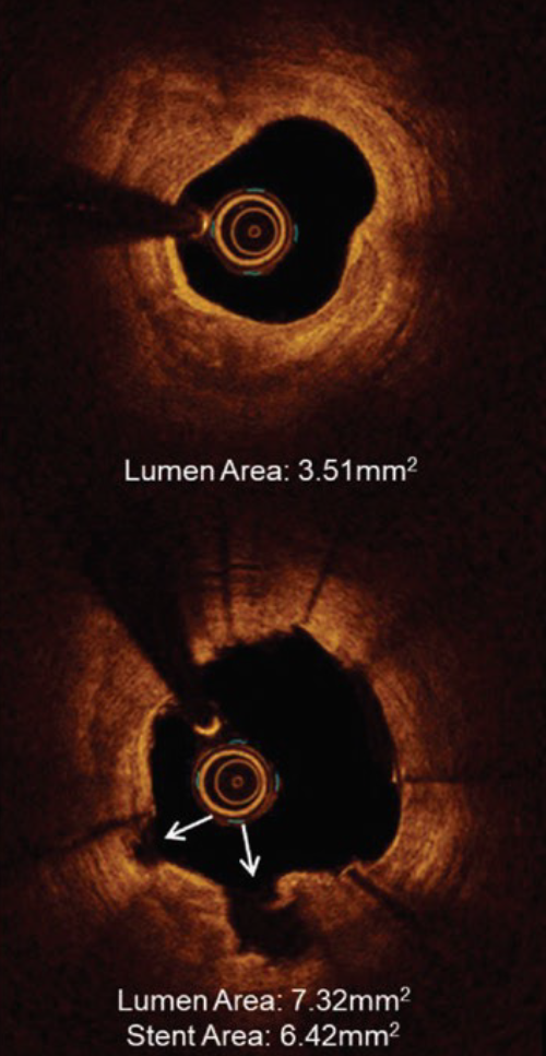
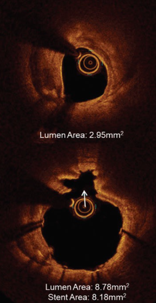
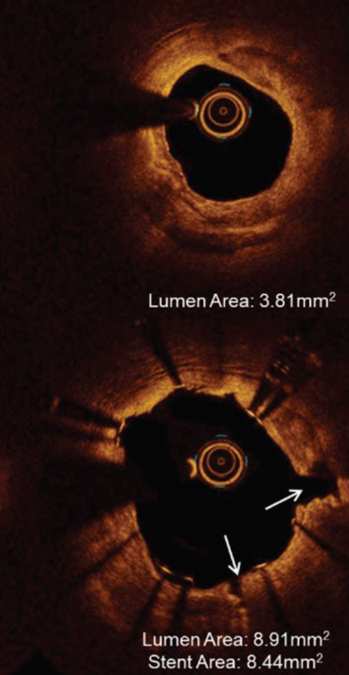
OCT Case Example

Proximal LAD with side branch involvement

Severe Ca++:
>270° & >1mm thick
Ca++ Fractures:
Multiple locations (white arrows)
Final Result:
Full stent expansion Large acute gain
A. Proximal vessel
cad ii case example a
B. Maximum Calcification
cad ii case example b
C. Distal vessel
cad ii case example c

Ready to Make Waves with Shockwave Medical?

Looking for careers? Click here.